Session Information
Session Type: Poster Session A
Session Time: 10:30AM-12:30PM
Background/Purpose: Evidence supports the benefits of exercise on joint health and its protective effects against osteoarthritis (OA) progression [Allen J, A&R, 2010]. However, most animal studies initiate exercise immediately after OA induction, failing to accurately reflect clinical OA progression, which was typically diagnosed at late or end-stage [Glyn-Jones S, Lancet, 2015]. Thus, the present study investigates the effects of treadmill exercise at different OA stages to optimize the timing of the intervention using mouse destabilization of the medial meniscus (DMM) model.
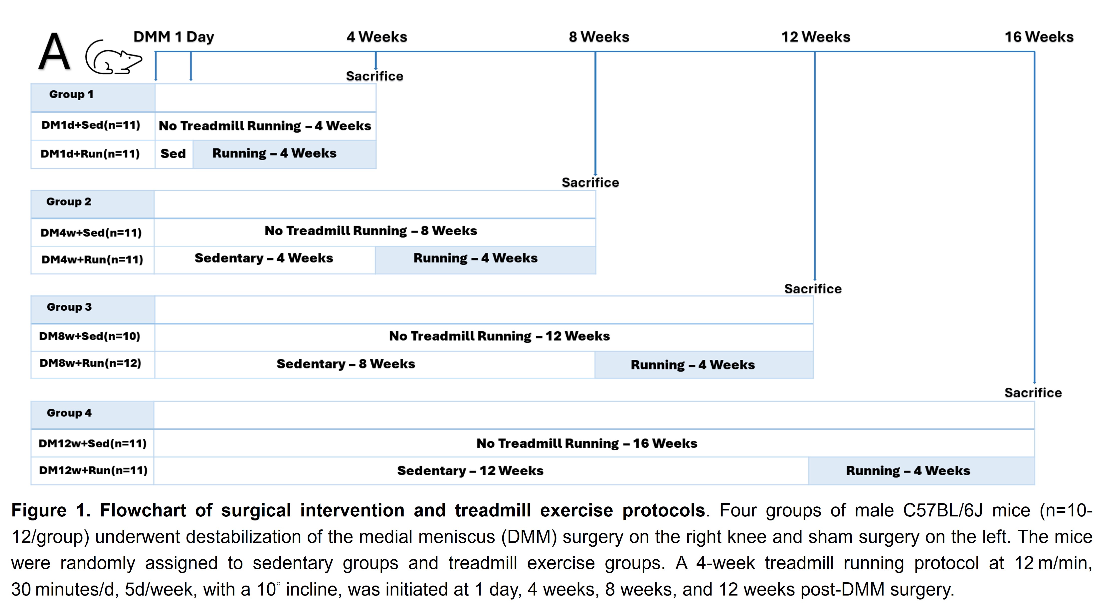
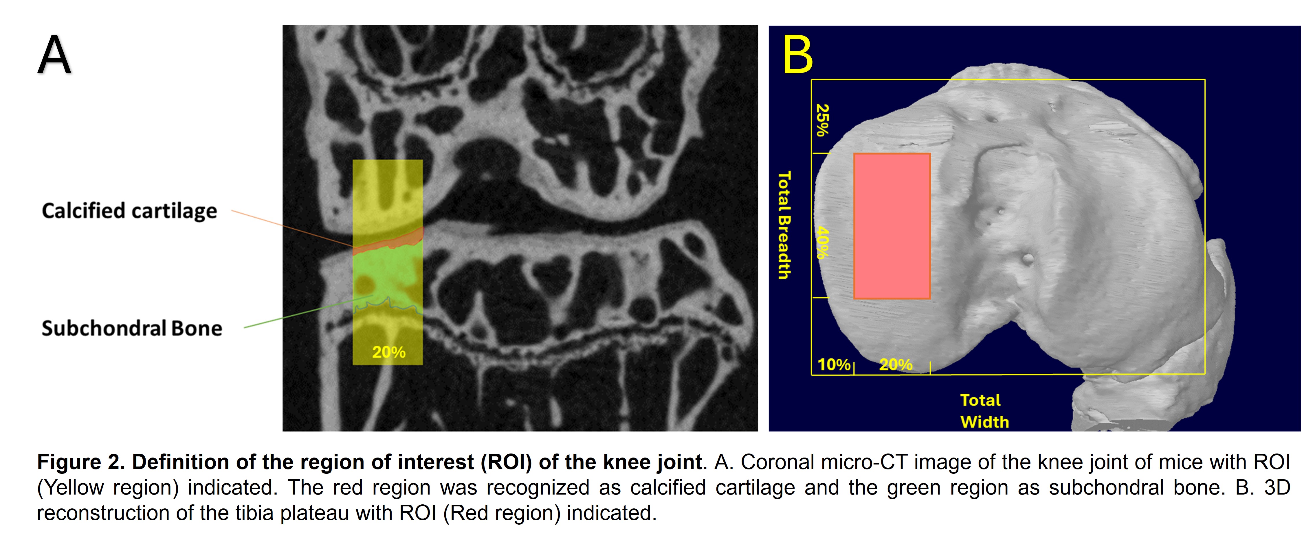
Methods: Four groups of male C57BL/6J mice (n=10-12/group) underwent either DMM or sham surgery were randomly assigned to sedentary or treadmill exercise groups. A 4-week treadmill running protocol (Fig. 1) was initiated at 1 day, 4 weeks, 8 weeks, and 12 weeks post-DMM surgery. Bilateral knee joints were collected at the end of the intervention, with left knee joints serving as sham. Micro-CT and Safranin O (SafO) staining were used to assess OARSI score, the thickness and cell density of articular cartilage (AC) and calcified cartilage (CC), and bone fraction and thickness of subchondral bone (SB) in medial tibia plateau (Fig. 2).
Results: Micro-CT analysis showed a decrease in SB thickness 4 weeks after DMM surgery yet an increase at 16 weeks post-operatively, while CC thickness was consistently reduced at all-time points after DMM surgery. Initiating treadmill exercise 1 day after DMM surgery resulted in improved SB BV/TV and thickness compared to sedentary mice. These benefits diminished when exercise started 4 weeks or later post-DMM surgery. Exercise commencing 8 weeks post-DMM surgery appeared detrimental, further reducing CC thickness. Exercise at 12 weeks post-DMM surgery exacerbated CC and SB deterioration. SafO staining revealed consistently elevated OARSI scores and decreased AC thickness and cell density. However, OA mitigation was observed when exercise began 1 day post-DMM surgery, with improved OARSI score and AC thickness. Exercise starting 4 weeks post-surgery potentially preserved AC thickness but reduced AC cell density. These parameters deteriorated when exercise began 8 weeks post-DMM surgery. After 12 weeks of DMM surgery, CC cell density further decreased following the exercise due to tremendous loss of AC.
Conclusion: Our data indicate a consistent decline in AC and CC properties during OA progression, while SB thickness decreased in early-stage OA but increased in end-stage OA. The transient decrease of SB thickness could be attributed to bone remodeling and is an important initiating factor in OA pathogenesis, which could not be detected clinically. Treadmill exercise significantly benefits joint health in early OA. However, the protective effects faded when exercise began 4 weeks post-DMM surgery, and destructive effects were noted when exercise was initiated 8 or 12 weeks after the surgery. These results indicate that regular exercise may not be suitable for late or end-stage OA. In summary, while regular exercise can be beneficial during early OA onset, its efficacy diminishes and potentially becomes harmful in advanced stages. An individualized exercise protocol is recommended for maximizing therapeutic efficacy in OA.
To cite this abstract in AMA style:
Chen M, Cui M, Yang Y, Wang B. The Impact of Treadmill Exercise on Various Stages of Osteoarthritis Progression in a Murine Osteoarthritis Model [abstract]. Arthritis Rheumatol. 2024; 76 (suppl 9). https://acrabstracts.org/abstract/the-impact-of-treadmill-exercise-on-various-stages-of-osteoarthritis-progression-in-a-murine-osteoarthritis-model/. Accessed .« Back to ACR Convergence 2024
ACR Meeting Abstracts - https://acrabstracts.org/abstract/the-impact-of-treadmill-exercise-on-various-stages-of-osteoarthritis-progression-in-a-murine-osteoarthritis-model/


