ACR Meeting Abstracts

ACR Meeting Abstracts

  • Meetings
    • ACR Convergence 2024
    • ACR Convergence 2023
    • 2023 ACR/ARP PRSYM
    • ACR Convergence 2022
    • ACR Convergence 2021
    • ACR Convergence 2020
    • 2020 ACR/ARP PRSYM
    • 2019 ACR/ARP Annual Meeting
    • 2018-2009 Meetings
    • Download Abstracts
  • Keyword Index
  • Advanced Search
  • Your Favorites
    • Favorites
    • Login
    • View and print all favorites
    • Clear all your favorites
  • ACR Meetings

Abstract Number: 1811

The Immune Map of Lupus Nephritis: A Spatially Resolved Kidney Proteomic Approach

Chen-Yu Lee1, Matthew Caleb Marlin2, Xiaoping Yang1, Alessandra Ida Celia3, Vasileios Morkotinis4, Richard Furie5, Jill Buyon6, Chaim Putterman7, Jennifer Barnas8, Kenneth Kalunian9, Peter Izmirly10, Betty Diamond11, Anne Davidson12, Diane Kamen13, Jeff Hodgin14, the Accelerating Medicines Partnership RA/SLE15, Judith James2, Michelle Petri16, Joel Guthridge2, Avi Rosenberg1 and Andrea Fava1, 1Johns Hopkins UNiversity, Baltimore, MD, 2Oklahoma Medical Research Foundation, Oklahoma City, OK, 3Sapienza University of Rome, Rome, Italy, 4University of Oklahoma Health Sciences Center, Oklahoma City, OK, 5Northwell Health, Manhasset, NY, 6NYU Grossman School of Medicine, New York, NY, 7Albert Einstein College of Medicine, Safed, Israel, 8University of Rochester, Rochester, NY, 9University of California San Diego, La Jolla, CA, 10New York University Grossman School of Medicine, New York, NY, 11The Feinstein Institutes for Medical Research, Manhasset, NY, 12Feinstein Institutes for Medical Research, Manhasset, NY, 13Medical University of South Carolina, Charleston, SC, 14University of Michigan, Michigan, 15multiple, multiple, 16Johns Hopkins University School of Medicine, Timonium, MD

Meeting: ACR Convergence 2024

Keywords: Bioinformatics, Biomarkers, Disease Activity, Lupus nephritis, Systemic lupus erythematosus (SLE)

  • Tweet
  • Click to email a link to a friend (Opens in new window) Email
  • Click to print (Opens in new window) Print
Session Information

Date: Monday, November 18, 2024

Title: SLE – Etiology & Pathogenesis Poster

Session Type: Poster Session C

Session Time: 10:30AM-12:30PM

Background/Purpose: Treatment response rates in lupus nephritis (LN) remain suboptimal, highlighting the need for a better understanding of LN pathogenesis to enhance treatment strategies. Single-cell transcriptomic studies are providing an unprecedented catalog of cell states in LN, yet their spatial organization is not well understood. Given that structure underlies function, our objective was to delineate the spatial organization of immune cells in LN.

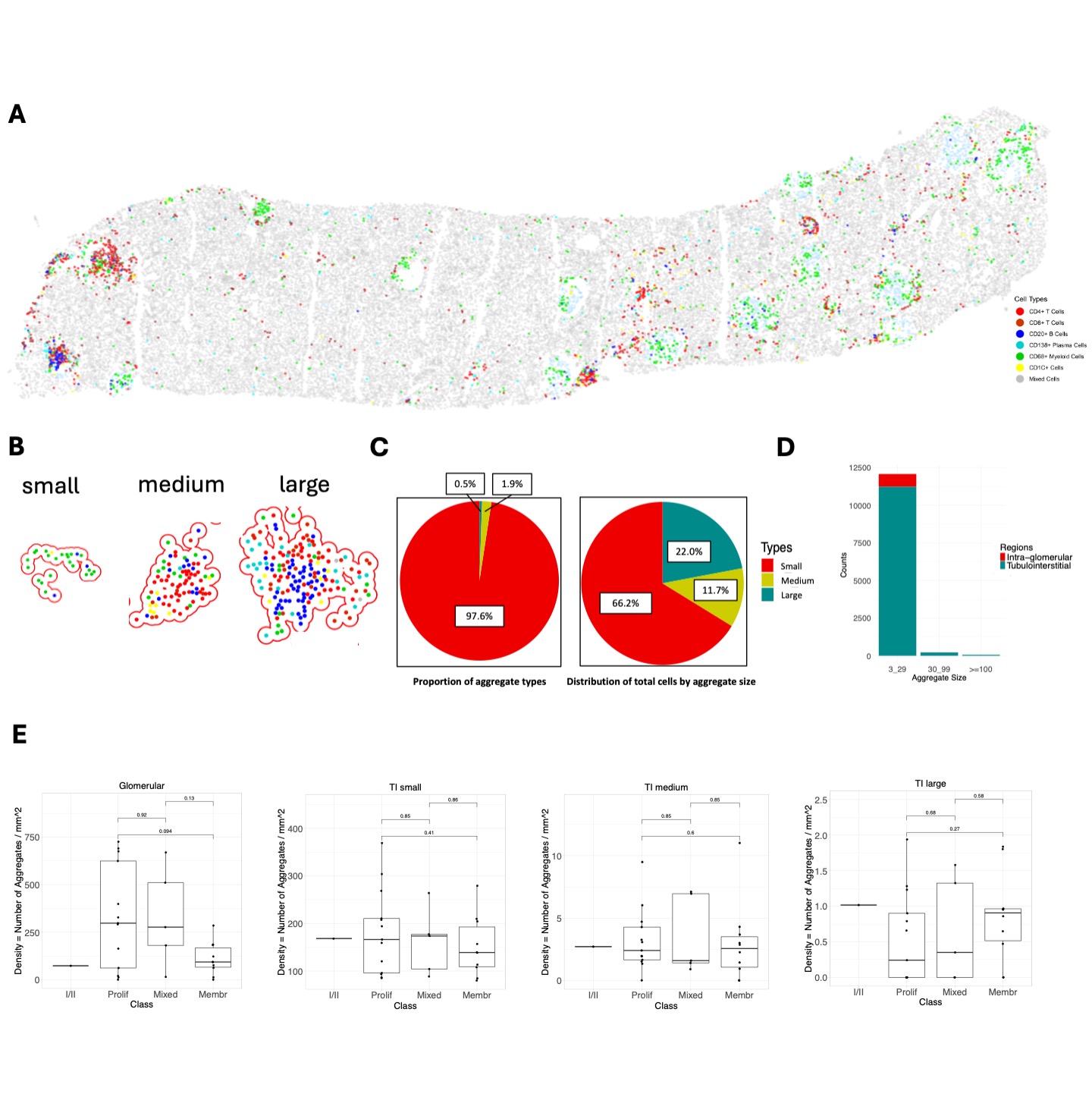
Methods: We developed a serial immunohistochemistry (sIHC) workflow (18-plex), consisting of cycles of staining, imaging, and de-staining. Image processing was carried out using HALO (Indica Labs), including AI-assisted tissue classification. Cell type annotation was performed at low resolution clustering for this preliminary analysis. Immune cell aggregates were defined using DBSCAN as a minimum of 3 cells within a radius (epsilon) of 50 μm to infer interactions between cells. Tubulointerstitial (TI) aggregate sizes were categorized into small (3-29 cells), medium (30-99 cells), and large ( >100 cells) based on the frequency distribution (Fig. 1A-B). Glomerular infiltrates were small and were not subdivided.

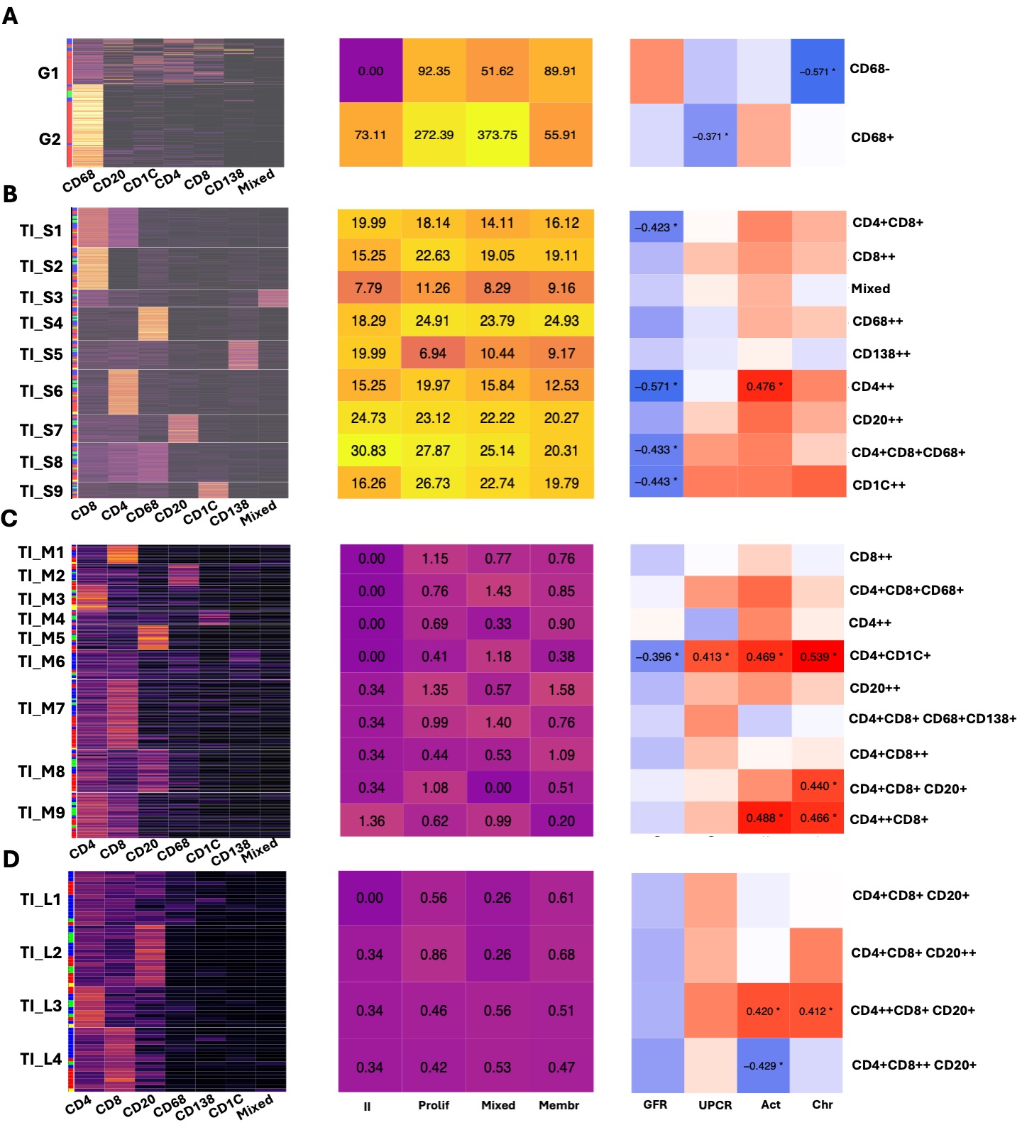
Results: In this analysis, we included 29 clinically indicated kidney biopsies classified as LN (13 pure proliferative, 10 pure membranous, 5 mixed, and 1 ISN class II) resulting in 1,913,845 cells (182,783 immune cells). We identified 12,371 cellular aggregates. Most (97%) aggregates were small (< 30 cells) (Fig. 1C-D), however, medium and large aggregates included 33.7% of immune cells. Overall, aggregate density was similar across LN classes, irrespective of size and region. Glomerular aggregates were small and primarily composed of CD68+ myeloid cells although some aggregates included T, B, dendritic and, occasionally, plasma cells (Fig. 2A).  Proliferative and mixed classes had more CD68-rich aggregates, but some were also found in pure membranous. Glomerular lymphocyte-rich aggregates negatively correlated with chronicity (Fig. 2A).  TI aggregate density was similar across LN classes (Fig. 1E) and negatively correlated with GFR. Significant heterogeneity in aggregate composition revealed >10 aggregate subtypes according to composition and size (Fig. 2). Small aggregates tended to be restricted to 1-2 cell types each, while medium and large aggregates included mixed proportions of CD4+ T, CD8+ T, B, dendritic, myeloid, and plasma cells likely representing germinal center-like structures (Fig. 2). Distinct TI aggregate subtypes associated with specific clinical and pathological features (Fig. 2B-C).


Conclusion: We describe the heterogeneity in glomerular and TI immune cell structures in LN, offering insights into LN pathological processes and potential cellular interactions based on proximity. Tubulointerstitial infiltrates appear similar in membranous and proliferative LN, yet specific immune structures are linked to distinct clinical and pathological features with many linked to worse GFR, regardless of class. Analysis of 90 additional biopsies is underway to validate and expand these findings.

Supporting image 1

Figure 1. Demographics of intrarenal immune cell aggregates. (A) Digitalized biopsy showing an example of the distribution of immune cells in LN. (B) Examples of intrarenal immune cell aggregates of different sizes. (C) Distribution of aggregates by size and by total cells. (D) Distribution of aggregates by size and region. (E) Density of aggregate types according to size and class.

Supporting image 2

Figure 2. Correlation between aggregate subtypes and clinical features. Heatmaps on the left displaying the subtypes of immune cell aggregates. The proportion of immune cell types in each aggregate was used for K-means clustering to determine aggregate subtypes. The column annotation rows of the heatmaps represent LN classes, red: proliferative; green: mixed, blue: pure membranous; yellow: ISN class II. Heatmaps in the middle show the average density of different aggregates, measured as the average number of aggregates per mm². Heatmaps on the right showing the correlation matrices between the aggregate subtypes and clinical features. Clinical features, including GFR, urinary protein-creatinine ratio (UPCR), and NIH activity and chronicity indices, were correlated with each aggregate subtype using Pearson’s correlation coefficient. Correlation coefficients with statistical significance are shown and marked with asterisks. (A) Glomerular small aggregate (B) Tubulointerstitial small aggregate (C) Tubulointerstitial medium aggregate (D) Tubulointerstitial large aggregate. Act: NIH activity index; Chr: NIH chronicity index


Disclosures: C. Lee: None; M. Marlin: None; X. Yang: None; A. Celia: None; V. Morkotinis: None; R. Furie: AstraZeneca, 1, 2, 6; J. Buyon: Artiva Biotherapeutics, 1, Bristol-Myers Squibb(BMS), 1, 2, Equillium, 1, GlaxoSmithKlein(GSK), 1, 2, Otsuka Pharmaceuticals, 1, Related Sciences, 1, 2; C. Putterman: Equillium, 2, Progentec, 2; J. Barnas: None; K. Kalunian: None; P. Izmirly: Hansoh Bio, 2; B. Diamond: adicet, 2, alpine, 12, dsmb, atara, 2, DBV, 2, icell, 2, sail, 2; A. Davidson: EMD Serono, 5; D. Kamen: Alpine Immune Sciences, 1, Bristol Myers Squibb (BMS), 1; J. Hodgin: None; t. Accelerating Medicines Partnership RA/SLE: None; J. James: GlaxoSmithKlein(GSK), 1, Progentec Diagnostics, Inc., 5, 10; M. Petri: Amgen, 2, AnaptysBio, 2, Annexon Bio, 2, Arthros-FocusMedEd, 6, AstraZeneca, 2, 5, Atara Biosciences, 2, Aurinia, 5, 6, Autolus, 2, Avoro Ventures, 2, Biocryst, 2, Boxer Capital, 2, Cabaletto Bio, 2, Caribou Biosciences, 2, CTI, 1, CVS Health, 1, Eli Lilly, 2, 5, Emergent Biosolutions, 1, Ermiium, 2, Escient Pharmaceuticals, 2, Exagen, 5, Exo Therapeutics, 2, Gentibio, 2, GlaxoSmithKlein(GSK), 2, 5, iCell Gene Therapeutics, 2, Innovaderm Research, 2, IQVIA, 1, Janssen, 5, Kira Pharmaceuticals, 2, Merck/EMD Serono, 1, Nexstone Immunology, 2, Nimbus Lakshmi, 2, Novartis, 2, PPD Development, 2, Precision Biosciences, 2, Proviant, 2, Regeneron Pharmaceuticals, 2, Sanofi, 2, Seismic Therapeutic, 2, Senti Bioscienes, 2, Sinomab Biosciences, 2, Takeda, 2, Tenet Medicines Inc, 2, TG Therapeutics, 2, UCB, 2, Vertex Pharmaceuticals, 2, Worldwide Clinical Trials, 1, Zydus, 2; J. Guthridge: AstraZeneca, 5, Bristol-Myers Squibb(BMS), 5; A. Rosenberg: None; A. Fava: Annexionbio, 2, Arctiva, 2, AstraZeneca, 2, Exagen, 5, Novartis, 6, UCB, 2.

To cite this abstract in AMA style:

Lee C, Marlin M, Yang X, Celia A, Morkotinis V, Furie R, Buyon J, Putterman C, Barnas J, Kalunian K, Izmirly P, Diamond B, Davidson A, Kamen D, Hodgin J, Accelerating Medicines Partnership RA/SLE t, James J, Petri M, Guthridge J, Rosenberg A, Fava A. The Immune Map of Lupus Nephritis: A Spatially Resolved Kidney Proteomic Approach [abstract]. Arthritis Rheumatol. 2024; 76 (suppl 9). https://acrabstracts.org/abstract/the-immune-map-of-lupus-nephritis-a-spatially-resolved-kidney-proteomic-approach/. Accessed .
  • Tweet
  • Click to email a link to a friend (Opens in new window) Email
  • Click to print (Opens in new window) Print

« Back to ACR Convergence 2024

ACR Meeting Abstracts - https://acrabstracts.org/abstract/the-immune-map-of-lupus-nephritis-a-spatially-resolved-kidney-proteomic-approach/

Advanced Search

Your Favorites

You can save and print a list of your favorite abstracts during your browser session by clicking the “Favorite” button at the bottom of any abstract. View your favorites »

All abstracts accepted to ACR Convergence are under media embargo once the ACR has notified presenters of their abstract’s acceptance. They may be presented at other meetings or published as manuscripts after this time but should not be discussed in non-scholarly venues or outlets. The following embargo policies are strictly enforced by the ACR.

Accepted abstracts are made available to the public online in advance of the meeting and are published in a special online supplement of our scientific journal, Arthritis & Rheumatology. Information contained in those abstracts may not be released until the abstracts appear online. In an exception to the media embargo, academic institutions, private organizations, and companies with products whose value may be influenced by information contained in an abstract may issue a press release to coincide with the availability of an ACR abstract on the ACR website. However, the ACR continues to require that information that goes beyond that contained in the abstract (e.g., discussion of the abstract done as part of editorial news coverage) is under media embargo until 10:00 AM ET on November 14, 2024. Journalists with access to embargoed information cannot release articles or editorial news coverage before this time. Editorial news coverage is considered original articles/videos developed by employed journalists to report facts, commentary, and subject matter expert quotes in a narrative form using a variety of sources (e.g., research, announcements, press releases, events, etc.).

Violation of this policy may result in the abstract being withdrawn from the meeting and other measures deemed appropriate. Authors are responsible for notifying colleagues, institutions, communications firms, and all other stakeholders related to the development or promotion of the abstract about this policy. If you have questions about the ACR abstract embargo policy, please contact ACR abstracts staff at [email protected].

Wiley

  • Online Journal
  • Privacy Policy
  • Permissions Policies
  • Cookie Preferences

© Copyright 2025 American College of Rheumatology