Session Information
Session Type: Poster Session B
Session Time: 9:00AM-11:00AM
Background/Purpose: While magnetic resonance imaging and ultrasound (US) imaging have been shown to be useful in the detection of subclinical joint inflammation in rheumatoid arthritis (RA), the literature on the use of thermal imaging (TI) in detecting subclinical disease in RA is presently lacking. The purpose of this RA study is to investigate whether there exists any correlation(s) between thermographic parameters and US-detected joint inflammation at clinically non-tender and non-swollen metacarpophalangeal joints (MCPJs).
Methods: RA patients (fulfilling the 2010 RA classification criteria) with clinically non-tender and non-swollen MCPJs underwent TI and US imaging of their 10 MCPJs on the same study visit. The thermographic parameters (maximum (Tmax), minimum (Tmin) and average (Tavg) temperatures in degree Celsius) were recorded from the dorsal surface of each MCPJ and summed up to obtain the respective Total Tmax, Total Tmin and Total Tavg per patient. Ultrasound power Doppler (PD) and grey-scale (GS) joint inflammation were scored semi-quantitatively (0-3) at the dorsal aspect of each MCPJ and summed up to obtain the respective Total PD and Total GS scores per patient. At each MCPJ, US synovitis is defined as PD ≥1 or GS ≥2 [1]. At the patient level, Pearson’s correlation coefficient was used to correlate Total Tmax, Total Tmin and Total Tavg with Total PD score, Total GS score and the number of joint(s) with US synovitis (PD ≥1 or GS ≥2). Among associations which were statistically significant, simple linear regression was applied to characterise relationship between variables.
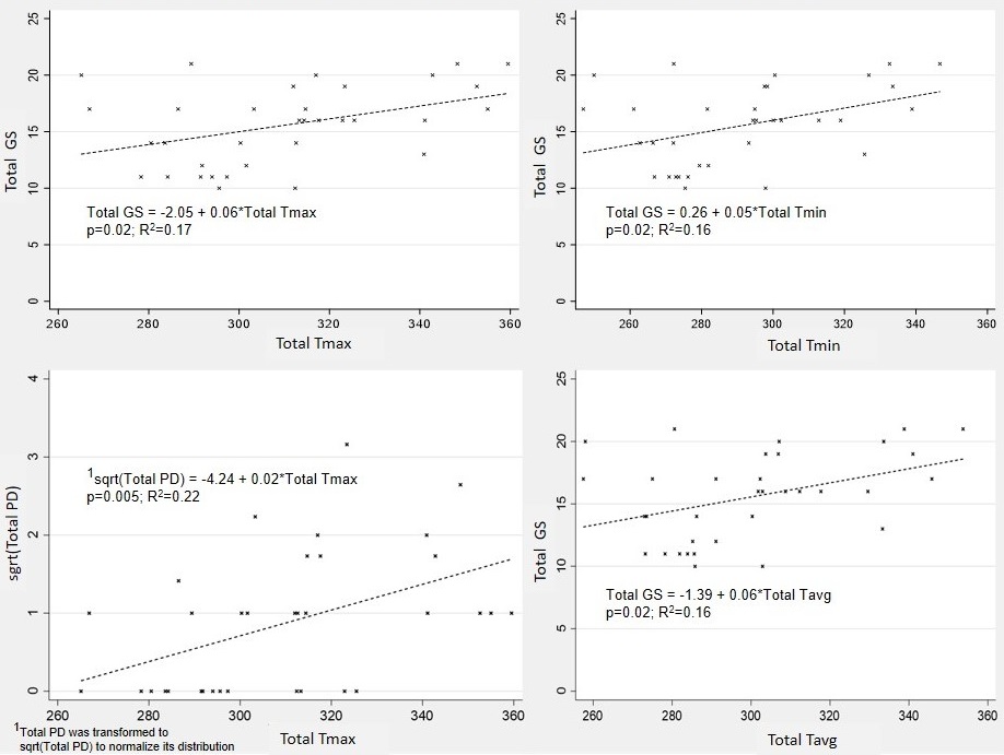
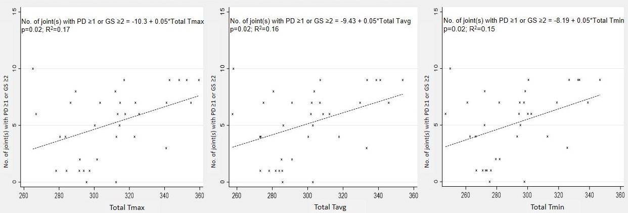
Results: A total of 340 MCPJs (clinically non-tender and non-swollen) were examined among 34 RA patients. In this cross-sectional study, the patients’ baseline characteristics are as follows: age, mean (SD) 58.3 (12.5) years; Chinese (n = 25, 73.5%); female (n = 30, 88.2%); DAS28, mean (SD) 3.13 (0.84); disease duration, mean (SD) 36.9 (58.1) months. Table 1 summarises the results from the correlation analysis. Total Tmax correlated significantly (P < 0.05) with Total PD score (r = 0.38), Total GS score (r = 0.41) and the number of joint(s) with PD ≥1 or GS ≥2 (r = 0.41). Total Tmin correlated significantly (P < 0.05) with Total GS score (r = 0.40) and the number of joint(s) with PD ≥1 or GS ≥2 (r = 0.39). Total Tavg correlated significantly (P < 0.05) with Total GS score (r = 0.41) and the number of joint(s) with PD ≥1 or GS ≥2 (r = 0.40).The relationship (only associations which were statistically significant are shown) of thermographic parameters with US variables and number of joint(s) with PD ≥1 or GS ≥2 are shown in Figure 1 and 2, respectively.
Conclusion: To the best of our knowledge, our RA study is the first to show that results from TI correlates significantly with US PD and GS joint inflammation as well as the number of joint(s) with ultrasound synovitis (PD ≥1 or GS ≥2) at the MCPJs. Like US imaging, TI can help detect subclinical joint inflammation at clinically non-tender and non-swollen MCPJs and further investigative work using TI in the detection of subclinical disease in RA appears warranted.
1. Hirata A, et al. Arthritis Care Res (Hoboken). 2017;69:801-806.
To cite this abstract in AMA style:
Tan Y, Lim G. The Detection of Subclinical Joint Inflammation by Thermal and Ultrasound Imaging at the Metacarpophalangeal Joints in Patients with Rheumatoid Arthritis [abstract]. Arthritis Rheumatol. 2023; 75 (suppl 9). https://acrabstracts.org/abstract/the-detection-of-subclinical-joint-inflammation-by-thermal-and-ultrasound-imaging-at-the-metacarpophalangeal-joints-in-patients-with-rheumatoid-arthritis/. Accessed .« Back to ACR Convergence 2023
ACR Meeting Abstracts - https://acrabstracts.org/abstract/the-detection-of-subclinical-joint-inflammation-by-thermal-and-ultrasound-imaging-at-the-metacarpophalangeal-joints-in-patients-with-rheumatoid-arthritis/


