Session Information
Date: Sunday, November 13, 2016
Title: Imaging of Rheumatic Diseases - Poster I: Ultrasound and Emerging Technologies
Session Type: ACR Poster Session A
Session Time: 9:00AM-11:00AM
Background/Purpose : Early treatment in enthesitis-related arthritis (ERA) may have a disease-modifying effect with consequently improved outcomes. However, disease activity may be difficult to assess, leading to a lack of confidence that inflammation is adequately controlled. Clinical evaluation is helpful in assessing disease activity of peripheral joints in ERA, but is less accurate for assessing sacroiliitis. Magnetic resonance imaging (MRI) is sensitive for detecting sacroiliitis, but current imaging methods are qualitative and rely on visual assessment by a radiologist. Quantitative imaging biomarkers (QIBs) rely on pixel values in the image itself, and may therefore be used to assess inflammation in a more objective, reproducible fashion. This study aims to evaluate diffusion-weighted imaging (DWI) as QIB of therapeutic response in adolescents with enthesitis-related arthropathy (ERA).
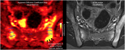
Methods : 22 adolescents with ERA underwent routine MRI and DWI before and after tumour necrosis factor inhibitor (TNFi) therapy [Figure 1]. Each patientÕs images were visually scored by two radiologists using a modification of the Spondyloarthritis Research Consortium of Canada (SPARCC) system (1). Sacroiliac joint apparent diffusion coefficient (ADC) and normalized ADC (nADC) were measured for each patient using a previously described technique (2). Therapeutic clinical response was defined as an improvement in physician global assessment of more than 30% and radiological response defined as at least a 2.5-point drop in SPARCC score.
Results: For both radiological and clinical definitions of response, reductions in ADC and nADC after treatment were greater in responders than in non-responders (for radiological response: ADC: p<0.01; nADC: p=0.055; for clinical response: ADC: p=0.33; nADC: p=0.089). ADC and nADC could predict radiological response with a high level of sensitivity and specificity (the area under the receiver operating characteristic curves were ADC: 0.97, nADC: 0.82).
Conclusion : DWI measurements reflect response to TNFi treatment in ERA patients with sacroiliitis. DWI is more objective than visual scoring, and has the potential to be automated. ADC/nADC could be used as biomarkers of sacroiliitis in the clinic and in clinical trials. References 1. Maksymowych WP et al. Arthritis Care Res. 2005;53:703Ð9. 2. Vendhan K et al. Br J Radiol. 2015;18:20150775. Figures
Figure 1 Ð Quantitative DWI (left) and conventional STIR imaging (right) in an ERA patient with sacroiliitis. Intensities in the ADC map represent the ADC for each pixel (see colour bar) but pixels in the STIR images have arbitrary values. SIJ ADC is measured using three linear regions of interest (ROIs, solid blue lines) placed on each sacroiliac joint; this is repeated for four consecutive slices. A further ÔreferenceÕ ROI is placed on sacral bone to normalize SIJ ADC for individual patients (dotted blue line).
To cite this abstract in AMA style:
Bray T, Vendhan K, Ambrose N, Atkinson D, Punwani S, Fisher C, Sen D, Ioannou Y, Hall-Craggs M. The Apparent Diffusion Coefficient As a Quantitative Imaging Biomarker of Therapeutic Response in Enthesitis-Related Arthritis [abstract]. Arthritis Rheumatol. 2016; 68 (suppl 10). https://acrabstracts.org/abstract/the-apparent-diffusion-coefficient-as-a-quantitative-imaging-biomarker-of-therapeutic-response-in-enthesitis-related-arthritis/. Accessed .« Back to 2016 ACR/ARHP Annual Meeting
ACR Meeting Abstracts - https://acrabstracts.org/abstract/the-apparent-diffusion-coefficient-as-a-quantitative-imaging-biomarker-of-therapeutic-response-in-enthesitis-related-arthritis/

