ACR Meeting Abstracts

ACR Meeting Abstracts

  • Meetings
    • ACR Convergence 2024
    • ACR Convergence 2023
    • 2023 ACR/ARP PRSYM
    • ACR Convergence 2022
    • ACR Convergence 2021
    • ACR Convergence 2020
    • 2020 ACR/ARP PRSYM
    • 2019 ACR/ARP Annual Meeting
    • 2018-2009 Meetings
    • Download Abstracts
  • Keyword Index
  • Advanced Search
  • Your Favorites
    • Favorites
    • Login
    • View and print all favorites
    • Clear all your favorites
  • ACR Meetings

Abstract Number: 1648

Systemic Sclerosis Auto-antibody Profiles Predict Interstitial Lung Disease Onset but Not Progression

Boyang Zheng1, Mianbo Wang 2, Marvin Fritzler 3, May Choi 3, Murray Baron 4 and Marie Hudson 5, 1Jewish General Hospital, McGill University, Montreal, QC, Canada, 2Lady Davis Institute, Jewish General Hospital, Montreal, QC, Canada, 3Cumming School of Medicine, University of Calgary, Calgary, AB, Canada, 4Jewish General Hospital, McGill University, Montreal, Canada, 5Jewish General Hospital, Lady Davis Institute for Medical Research, and Department of Medicine, McGill University, Montreal, QC, Canada

Meeting: 2019 ACR/ARP Annual Meeting

Keywords: autoantibodies, interstitial lung disease, prognostic factors and pulmonary fibrosis, Systemic sclerosis

  • Tweet
  • Click to email a link to a friend (Opens in new window) Email
  • Click to print (Opens in new window) Print
Session Information

Date: Monday, November 11, 2019

Title: Systemic Sclerosis & Related Disorders – Clinical Poster II

Session Type: Poster Session (Monday)

Session Time: 9:00AM-11:00AM

Background/Purpose: Interstitial lung disease (ILD) is frequent manifestation and a leading cause of mortality in systemic sclerosis (SSc). Some SSc-related autoantibodies (aAbs), especially anti-topoisomerase I (ATA), are associated with an increased prevalence of ILD. In contrast, anti-centromere (ACA) may be protective. The association with other aAbs is unclear and it is unknown whether aAb profiles can predict the rate of ILD progression. Our goal was to examine the incidence of SSc-ILD and its subsequent progression between different aAb profiles. We hypothesized that while aAbs predict ILD development, all SSc-ILD progress similarly.

Methods: SSc subjects without pre-existing ILD in a large multi-center longitudinal cohort from 2004-2018 were included. ILD was defined by the presence of high-resolution computed tomography (HRCT) findings, or if no HRCT was available, then either a chest x-ray showing interstitial fibrosis, and/or the presence of “velcro like crackles” on exam. Clinically meaningful progression of ILD was defined using OMERACT criteria(1) of ≥10% relative decline in FVC, or ≥5% to < 10% relative decline in FVC and ≥15% relative decline in DLCO. Subjects were grouped based on single specificity aAbs: ATA, ACA, anti-RNA polymerase III (ARA), anti-PM/Scl, anti-Ku, anti-Th/To (hPOP1), anti-fibrillarin (AFA), or negative aAbs. The time to ILD onset was compared between groups using Kaplan Meier and multivariate Cox analyses adjusted for age, gender, diffuse (vs. limited) subset, physician global assessment of severity, immunosuppression exposure at or prior to baseline and presence of anti-Ro52/TRIM21 antibodies. In those with incident ILD, the time to clinically meaningful progression and mortality after ILD onset were examined. ATA served as the reference for all comparisons.

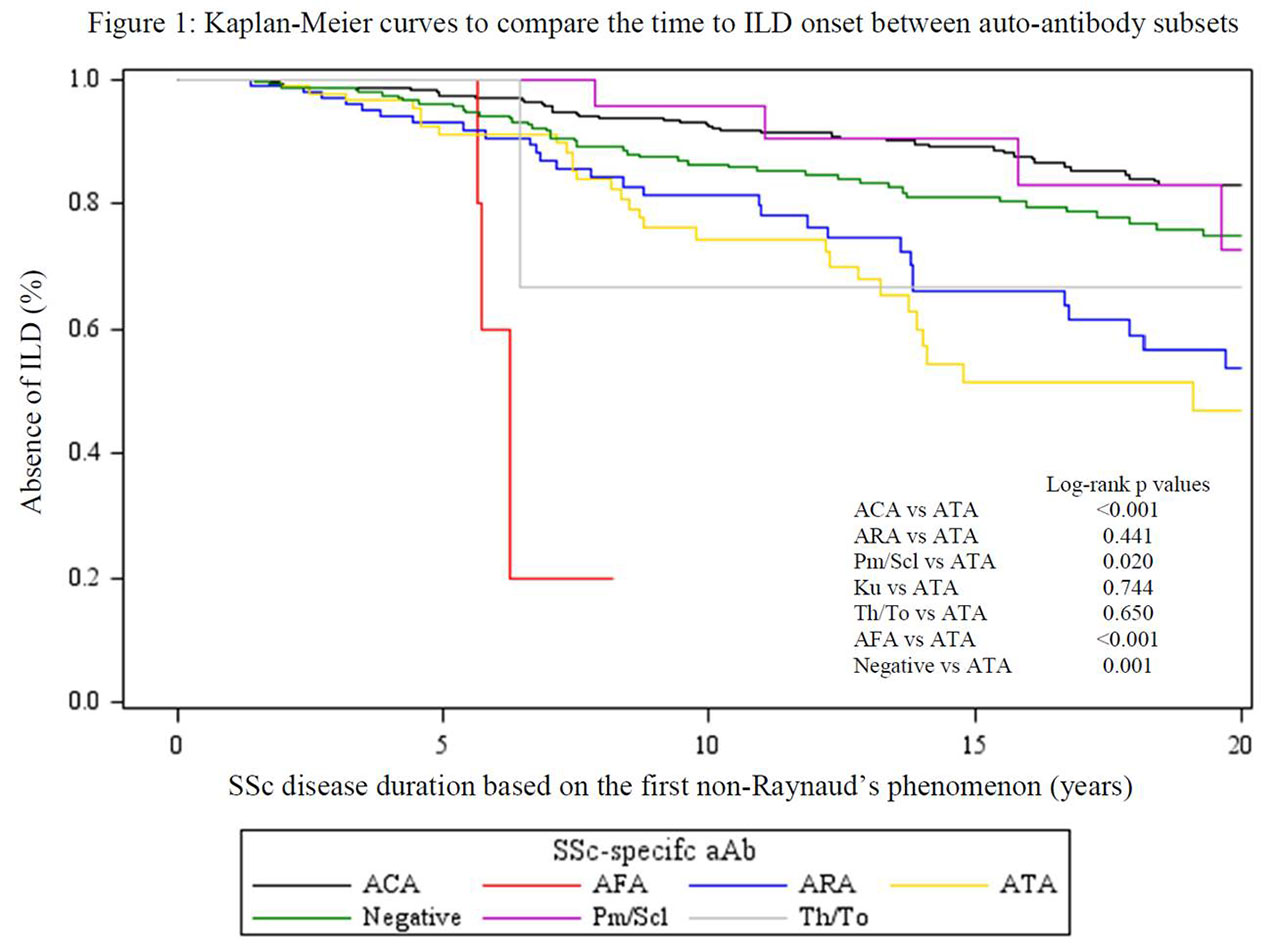
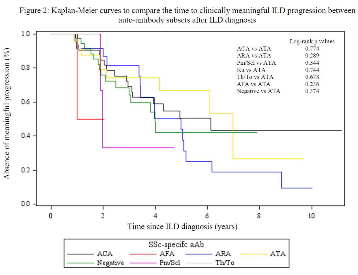
Results: Of 931 patients, 190 (20%) developed ILD: 60/429 (14%) ACA, 31/94 (33%) ATA, 32/114 (28%) ARA, 6/24 (25%) anti-Pm/Scl, 0/2 anti-Ku, 2/5 (40%) anti-Th/To, 4/5 (80%) AFA, and 55/258 (21%) negative aAb. Subjects with ATA developed ILD earlier than ACA, anti-PM/Scl and negative aAb groups (log rank p < 0.01, p = 0.02 and p < 0.01, respectively), although AFA group had the most rapid onset (p < 0.01) (Figure 1). In multivariate analyses, ACA and negative aAb groups were less likely (HR (95% CI) 0.36 (0.22, 0.57) and 0.53 (0.34, 0.84) respectively), whereas AFA was more likely (HR (95% CI) 4.12 (1.22, 13.86)) to develop ILD compared to ATA.  On the other hand, no differences in the time to clinically meaningful ILD progression (Figure 2) or survival after ILD onset were found between groups.

Conclusion: Compared to patients with ATA, AFA conferred a higher risk of developing ILD, whereas ACA, anti-PM/Scl and the absence of SSc specific aAbs were associated with a lower risk. Nevertheless, ILD developed in all aAb sub-groups and, once present, the aAb profile was not associated with ILD progression or subsequent mortality.

  1. Khanna D, Mittoo S, Aggarwal R, Proudman SM, Dalbeth N, Matteson EL, et al. Connective Tissue Disease-associated Interstitial Lung Diseases (CTD-ILD) – Report from OMERACT CTD-ILD Working Group. The Journal of rheumatology. 2015;42(11):2168-71.


Disclosure: B. Zheng, None; M. Wang, None; M. Fritzler, Alexion Canada, 7, BioRad, 5, Dr. Fooke Laboratorien GmbH, 5, Euroimmun GmbH, 5, 7, ImmunoConcepts, 7, Inova Diagnostics, 5, 7, 8, Inova Diagnostics Inc. San diego, CA, 5, Inova Dx, Mikrogen GmbH, 5, Werfen International, 5; M. Choi, None; M. Baron, None; M. Hudson, None.

To cite this abstract in AMA style:

Zheng B, Wang M, Fritzler M, Choi M, Baron M, Hudson M. Systemic Sclerosis Auto-antibody Profiles Predict Interstitial Lung Disease Onset but Not Progression [abstract]. Arthritis Rheumatol. 2019; 71 (suppl 10). https://acrabstracts.org/abstract/systemic-sclerosis-auto-antibody-profiles-predict-interstitial-lung-disease-onset-but-not-progression/. Accessed .
  • Tweet
  • Click to email a link to a friend (Opens in new window) Email
  • Click to print (Opens in new window) Print

« Back to 2019 ACR/ARP Annual Meeting

ACR Meeting Abstracts - https://acrabstracts.org/abstract/systemic-sclerosis-auto-antibody-profiles-predict-interstitial-lung-disease-onset-but-not-progression/

Advanced Search

Your Favorites

You can save and print a list of your favorite abstracts during your browser session by clicking the “Favorite” button at the bottom of any abstract. View your favorites »

All abstracts accepted to ACR Convergence are under media embargo once the ACR has notified presenters of their abstract’s acceptance. They may be presented at other meetings or published as manuscripts after this time but should not be discussed in non-scholarly venues or outlets. The following embargo policies are strictly enforced by the ACR.

Accepted abstracts are made available to the public online in advance of the meeting and are published in a special online supplement of our scientific journal, Arthritis & Rheumatology. Information contained in those abstracts may not be released until the abstracts appear online. In an exception to the media embargo, academic institutions, private organizations, and companies with products whose value may be influenced by information contained in an abstract may issue a press release to coincide with the availability of an ACR abstract on the ACR website. However, the ACR continues to require that information that goes beyond that contained in the abstract (e.g., discussion of the abstract done as part of editorial news coverage) is under media embargo until 10:00 AM ET on November 14, 2024. Journalists with access to embargoed information cannot release articles or editorial news coverage before this time. Editorial news coverage is considered original articles/videos developed by employed journalists to report facts, commentary, and subject matter expert quotes in a narrative form using a variety of sources (e.g., research, announcements, press releases, events, etc.).

Violation of this policy may result in the abstract being withdrawn from the meeting and other measures deemed appropriate. Authors are responsible for notifying colleagues, institutions, communications firms, and all other stakeholders related to the development or promotion of the abstract about this policy. If you have questions about the ACR abstract embargo policy, please contact ACR abstracts staff at [email protected].

Wiley

  • Online Journal
  • Privacy Policy
  • Permissions Policies
  • Cookie Preferences

© Copyright 2025 American College of Rheumatology