Session Information
Session Type: ACR Poster Session C
Session Time: 9:00AM-11:00AM
Background/Purpose: To examine whether synovial changes on ultrasound (US) associate with knee pain (KP) and/or underlying structural radiographic changes of osteoarthritis (OA).
Methods: In this case-control study, participants with early KP and established KP (more than 3 years duration) were compared with participants without KP. Participants were selected from the Knee Pain and Related Health in the Community (KPIC, n=9514) survey in Nottingham, UK. Cases and controls were age and gender matched. KP was defined as pain in or around the knee on most days for at least a month. Synovial changes (effusion, hypertrophy and Power Doppler (PD) signal) were measured by the two observers (inter-observer concordance correlation was 0.8 (0.6 to 0.9) for effusion and 0.7 (0.5 to 0.9) for synovial hypertrophy). OA structural changes were measured by standardised radiographs (semi-flexed weight-bearing and flexed skyline views) using the Nottingham Line Drawing Atlas (NLDA). Odds ratio (OR) and 95% confidence interval (CI) were estimated with adjustment for age, gender, body mass index and the total NLDA scores. Multinomial logistic regression was used to handle the case control study with three groups and to estimate ORs between early KP, established KP and no KP groups. A subgroup analysis in participants with unilateral KP was performed using a multilevel generalized linear mixed regression to compare painful knees with pain-free knees within persons (a well matched case control analysis).
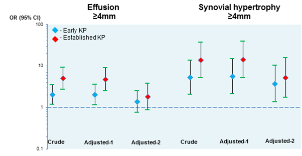
Results: A total of 420 participants were included of which 219 had early KP, 103 established KP and 98 no KP (mean age 60 years; 60% women). The prevalence of US effusion (≥4mm) were 24.5%, 39.8% and 62.1% in no KP, early KP and established KP. The prevalence of US hypertrophy (≥4mm) were 5.1%, 22.2% and 42.7% and the prevalence of PD signal were 0%, 3.2%, and 1.9% in these three groups. US effusion (≥4mm) increased the risk of early KP (OR 2.03, 95% CI 1.16 to 3.56) and established KP (4.75, 2.49 to 9.06). Similarly, hypertrophy (≥4mm) also increased the risk of early KP (5.63, 2.12 to 15.01) and established KP (14.24, 5.12 to 39.60). However, the association with effusion was diminished when adjusted for radiographic changes (Figure 1). In contrast, radiographic severity of OA on the NLDA was positively associated with KP, irrespective of the adjustment for the US features and other confounding factors.
Further analysis in people with unilateral KP (n=147) showed that the associations with both effusion and hypertrophy were diminished after adjusting for radiographic severity (1.02 (0.61 to 1.69) and 1.64 (0.85 to 3.17), respectively).
Conclusion: This study confirms that the association between US synovial changes and KP depends on the radiographic severity of OA, suggesting that US features reflect part of the overall structural changes occurring in OA joints and on their own are unlikely to be independent predictors of knee pain.
Figure 1. Association between US features and knee pain. Note that y axis is logarithmically scaled.
Abbreviations: OR – odds ratio; CI –confidence interval;
Adjusted-1 – adjusted for age, gender and BMI;
Adjusted-2 – adjusted for age, gender and BMI and global x-ray score.
To cite this abstract in AMA style:
Sarmanova A, Hall M, Fernandes G, Bhattacharya A, Valdes A, Walsh D, Doherty M, Zhang W. Synovial Changes Detected By Ultrasound and Its Association with Knee Pain: A Population-Based Case Control Study [abstract]. Arthritis Rheumatol. 2016; 68 (suppl 10). https://acrabstracts.org/abstract/synovial-changes-detected-by-ultrasound-and-its-association-with-knee-pain-a-population-based-case-control-study/. Accessed .« Back to 2016 ACR/ARHP Annual Meeting
ACR Meeting Abstracts - https://acrabstracts.org/abstract/synovial-changes-detected-by-ultrasound-and-its-association-with-knee-pain-a-population-based-case-control-study/

