Session Information
Date: Monday, November 6, 2017
Title: Systemic Lupus Erythematosus – Human Etiology and Pathogenesis Poster I
Session Type: ACR Poster Session B
Session Time: 9:00AM-11:00AM
Background/Purpose: Follicular helper T cells (Tfh) cells regulate the germinal center (GC) response by delivery of contact-dependent interactions and cytokines including IL-4, IFN-γ and IL-21. JAK-STAT signaling plays an important role in regulating these cytokines associated biological responses. Signal Transducer and Activator of Transcription proteins (STATs) are intracellular transcriptional factors that are activated and recruited to the cytokine receptors through phosphorylation by Janus Kinase (JAK).
There are several STATs (STAT1, STAT2, STAT3, STAT4, STAT5 and STAT6) in mammalian cells. STAT4 is the only STAT that demonstrates the genetic association to disease susceptibility in human systemic lupus by a number of Genome-Wide Association Studies. These genetic associations lie in the single-nucleotide polymorphisms (SNPs) among the noncoding regions. STAT4 SNP at rs7574865, which locates intron 3, is associated with increased sensitivity to IFNa signaling and gene expression, and suggests that these genetic variants may affect disease susceptibility through the gene’s transcription.
Role of STAT4 in disease pathology is still unclear. STAT-4 in human SLE has not been robustly studied.
Methods: B6.Sle1.Yaa mice were sacrificed at 2,4, and 6 months of age flow cytometry plots of Tfh cells or Th1 cells were obtained using FLOW. Flow cytometry plots of intracellular staining for the transcription factors Bcl6 and T-bet in Tfh cells over time were also performed. 40 ml peripheral blood was drawn during the clinical visit. Serum was separated and stored in -80 C. Mononuclear cells from the peripheral blood was isolated using Ficoll-Paque, suspended in serum with 10% DMSO and store in -80 degree for STAT4 assay.
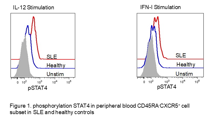
Results: In young lupus mice, Tfh cells co-express the Tfh and Th1 cell transcription factors Bcl6 and T-bet, respectively; with a decline of both in Tfh cells as the disease progresses. However, Tfh cells continue to co-produce similar levels of both IL-21 and IFN-γ during the progression of murine lupus. Transcriptional analysis of lupus-Tfh cells at different stages of disease revealed an increased STAT4 gene signature as the disease progresses. Moreover, we observed that lupus-Tfh cells continue to phosphorylate STAT4 (pSTAT4) as the disease worsens, consistent with the prolonged production of pathogenic IL-21 and IFN-γ. In the blood of lupus patients, activated (CD4+CD45RA–) T cells also secrete IL-21 and IFN-γ. STAT4 activity was measured in these cells using FLOW. Unstimulated CD4+CD45RA– T cells from lupus patients had an increase in pSTAT4 compared to that of healthy controls. Furthermore, pSTAT4 was further increased with IFN-a or IL-12 stimulation in the lupus patients compared to controls.
Conclusion: In SLE, T-bet and Bcl6 expression decline in Tfh cells, while increased STAT4-guided expression appears to drive pathogenic cytokine production, providing a potential therapeutic target for SLE.
To cite this abstract in AMA style:
Koumpouras F, Dong X, Weinstein J, Craft JE. STAT4 Regulates Pathogenic IL-21 and IFN-γ in Tfh Cells in Murine and Human Lupus [abstract]. Arthritis Rheumatol. 2017; 69 (suppl 10). https://acrabstracts.org/abstract/stat4-regulates-pathogenic-il-21-and-ifn-%ce%b3-in-tfh-cells-in-murine-and-human-lupus/. Accessed .« Back to 2017 ACR/ARHP Annual Meeting
ACR Meeting Abstracts - https://acrabstracts.org/abstract/stat4-regulates-pathogenic-il-21-and-ifn-%ce%b3-in-tfh-cells-in-murine-and-human-lupus/

