ACR Meeting Abstracts

ACR Meeting Abstracts

  • Meetings
    • ACR Convergence 2024
    • ACR Convergence 2023
    • 2023 ACR/ARP PRSYM
    • ACR Convergence 2022
    • ACR Convergence 2021
    • ACR Convergence 2020
    • 2020 ACR/ARP PRSYM
    • 2019 ACR/ARP Annual Meeting
    • 2018-2009 Meetings
    • Download Abstracts
  • Keyword Index
  • Advanced Search
  • Your Favorites
    • Favorites
    • Login
    • View and print all favorites
    • Clear all your favorites
  • ACR Meetings

Abstract Number: 0832

Spatial Transcriptomic Assessment of Histologically Damaged and Unaffected Glomeruli in Class III Pediatric Lupus Nephritis Reveals Distinct Transcriptional Programs

Sarah McCuaig1, Portia Kreiger1 and Edward Behrens2, 1Children's Hospital of Philadelphia, Philadelphia, PA, 2Children's Hospital of Philadelphia, West Chester, PA

Meeting: ACR Convergence 2024

Keywords: Lupus nephritis, Pediatric rheumatology, Systemic lupus erythematosus (SLE)

  • Tweet
  • Click to email a link to a friend (Opens in new window) Email
  • Click to print (Opens in new window) Print
Session Information

Date: Saturday, November 16, 2024

Title: Abstracts: Pediatric Rheumatology – Basic Science

Session Type: Abstract Session

Session Time: 3:00PM-4:30PM

Background/Purpose: Lupus nephritis (LN) occurs in over 50% of patients with pediatric systemic lupus erythematosus (pSLE) and results in significant morbidity due to suboptimal kidney remission rates and the sequelae of prolonged intensive immunosuppressive therapy. While the proximal drivers of LN remain obscure, Type I interferon (IFN) has often been invoked in pathogenesis. LN is often patchy, with some glomeruli severely damaged while others remain histologically unaffected in the same kidney. Yet, the factors that drive these patterns of local injury are unclear. Using novel spatial transcriptomic technology, we interrogated microanatomic transcriptional differences between histologically damaged and unaffected glomeruli in pSLE LN to understand local drivers of glomerular damage.

Methods: Archived pre-treatment FFPE pediatric renal biopsies were stained with H&E. Using the Visium 10x platform, spatially barcoded gene expression libraries mapped to 55μM diameter spots were sequenced.  A pediatric pathologist identified histologically damaged and unaffected glomeruli based on H&E staining. Loupe Browser (10X Genomics) was used to annotate damaged versus unaffected glomeruli and associated barcoded spots. Differential gene expression analysis of spots was performed in R. 2 cases of pSLE Class III LN, 2 cases of non-SLE glomerulonephritis (GN, C3 glomerulopathy and post-streptococcal GN), and 2 “healthy control” kidneys resected for non-immune pathology were assessed.

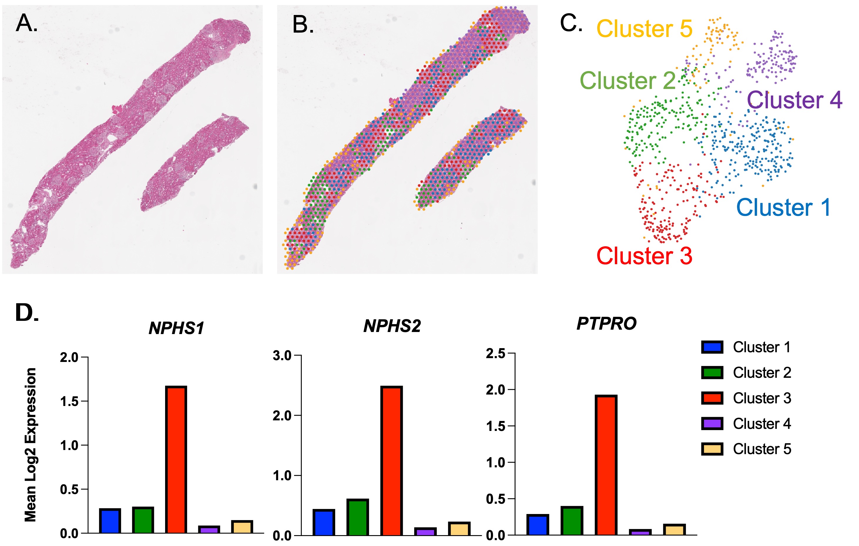
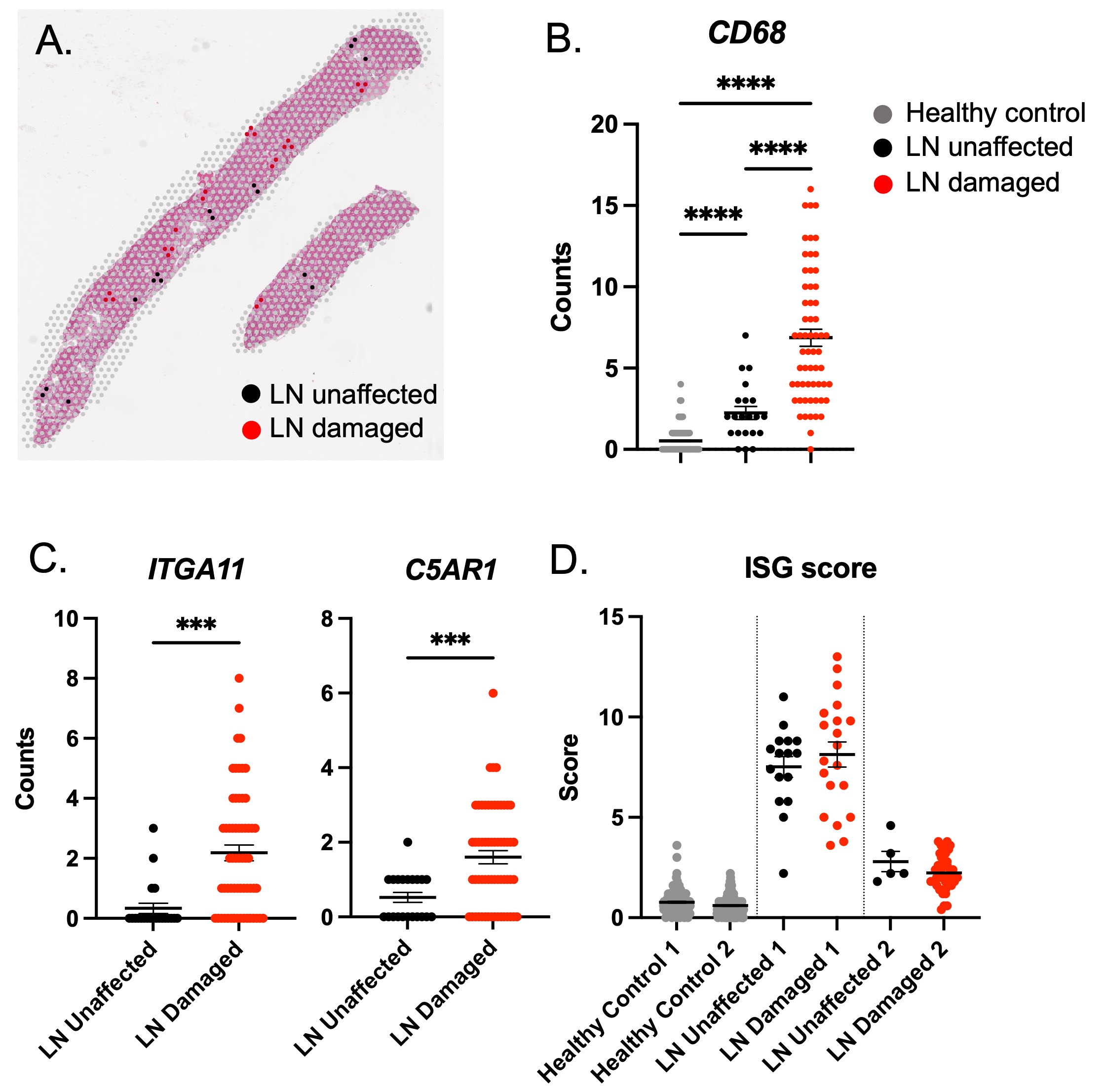
Results: High quality RNA was obtained from each specimen. Unsupervised clustering of spots identified glomeruli transcriptionally with excellent concordance to their microanatomic location (Figure 1). Differential gene expression analysis confirmed that pSLE glomeruli are transcriptionally distinct from non-SLE GN glomeruli and healthy control glomeruli. Moreover, among pSLE Class III LN glomeruli, damaged and unaffected glomeruli have unique transcriptional signatures. Predictably, damaged LN glomeruli have increased expression of the myeloid marker CD68 compared to unaffected LN glomeruli and healthy control glomeruli (Figure 2B). Damaged LN glomeruli also have increased expression of C5AR1 and ITGA11 compared to unaffected glomeruli, implying complement component 5a involvement and myofibroblast/fibrosis respectively (Figure 2C). Interestingly, while IFN stimulated gene (ISG) expression is elevated in pSLE compared to healthy control glomeruli, ISG expression is no different between damaged and unaffected LN glomeruli from the same kidney (Figure 2D).

Conclusion: Despite being canonically thought of as a disease of Type I IFN, IFN gene response does not associate with local glomerular damage in pSLE.  Rather, non-IFN related transcriptional events associate with damage including C5aR and ITGA11. These results suggest novel useful biomarkers and potential therapeutic targets for LN (eg. avacopan to target C5aR). Resolution of the local transcriptional events associated with injury in LN compared to non-SLE GN reveals novel biology that will inform future mechanistic studies using animal models, and translational clinical studies by pointing to novel targets.

Supporting image 1

Figure 1 – Unbiased graph-based clustering identifies glomeruli transcriptionally that corresponds to microanatomic location in pediatric lupus nephritis renal biopsies. Spatial transcriptomics using the Visium 10x platform was performed on FFPE renal biopsies from pediatric patients with LN (N=2), C3G (N=1), PSGN (N=1), and non-immune pathology “healthy control” (N=2). (A) Representative H&E section from LN biopsy. (B) Overlay of 55uM spots colored coded based on unbiased graph-based clustering. (C) UMAP projection of graph-based clusters. (D) Cluster 3 is defined by high expression of glomerular genes NPHS1, NPHS2, PTPRO. Accordingly, cluster 3 spatially correlates with glomeruli as seen in (B). Values are mean normalized gene UMI counts in the cluster.

Supporting image 2

Figure 2 – Transcriptional differences between histologically damaged and unaffected glomeruli in Class III pSLE GN. (A) Annotation of spots overlying glomeruli as either healthy or diseased by a pathologist using H&E histology. (B) Myeloid cell marker, CD68 expression in healthy control, LN unaffected, and LN damaged glomeruli. (C) Examples of genes found to be significantly upregulated in LN damaged compared to unaffected glomeruli. ITGA11 is integrin alpha 11, a gene associated with fibrosis. C5AR1 is the complement 5a receptor. (D) Interferon score, the average of the expression of IFI44, IFI44L, MX1, IFIT1, and HERC6, comparing healthy control to unaffected and damaged LN glomeruli stratified by patient. Each dot represents expression from a 55uM spot. N=2 pSLE Class III LN patients. N=2 healthy control patients.


Disclosures: S. McCuaig: None; P. Kreiger: AB2Bio, 5, Pharming, 2, Sobi, 2; E. Behrens: Ab2Bio, 5, Pharming, 2, Sobi, 2.

To cite this abstract in AMA style:

McCuaig S, Kreiger P, Behrens E. Spatial Transcriptomic Assessment of Histologically Damaged and Unaffected Glomeruli in Class III Pediatric Lupus Nephritis Reveals Distinct Transcriptional Programs [abstract]. Arthritis Rheumatol. 2024; 76 (suppl 9). https://acrabstracts.org/abstract/spatial-transcriptomic-assessment-of-histologically-damaged-and-unaffected-glomeruli-in-class-iii-pediatric-lupus-nephritis-reveals-distinct-transcriptional-programs/. Accessed .
  • Tweet
  • Click to email a link to a friend (Opens in new window) Email
  • Click to print (Opens in new window) Print

« Back to ACR Convergence 2024

ACR Meeting Abstracts - https://acrabstracts.org/abstract/spatial-transcriptomic-assessment-of-histologically-damaged-and-unaffected-glomeruli-in-class-iii-pediatric-lupus-nephritis-reveals-distinct-transcriptional-programs/

Advanced Search

Your Favorites

You can save and print a list of your favorite abstracts during your browser session by clicking the “Favorite” button at the bottom of any abstract. View your favorites »

All abstracts accepted to ACR Convergence are under media embargo once the ACR has notified presenters of their abstract’s acceptance. They may be presented at other meetings or published as manuscripts after this time but should not be discussed in non-scholarly venues or outlets. The following embargo policies are strictly enforced by the ACR.

Accepted abstracts are made available to the public online in advance of the meeting and are published in a special online supplement of our scientific journal, Arthritis & Rheumatology. Information contained in those abstracts may not be released until the abstracts appear online. In an exception to the media embargo, academic institutions, private organizations, and companies with products whose value may be influenced by information contained in an abstract may issue a press release to coincide with the availability of an ACR abstract on the ACR website. However, the ACR continues to require that information that goes beyond that contained in the abstract (e.g., discussion of the abstract done as part of editorial news coverage) is under media embargo until 10:00 AM ET on November 14, 2024. Journalists with access to embargoed information cannot release articles or editorial news coverage before this time. Editorial news coverage is considered original articles/videos developed by employed journalists to report facts, commentary, and subject matter expert quotes in a narrative form using a variety of sources (e.g., research, announcements, press releases, events, etc.).

Violation of this policy may result in the abstract being withdrawn from the meeting and other measures deemed appropriate. Authors are responsible for notifying colleagues, institutions, communications firms, and all other stakeholders related to the development or promotion of the abstract about this policy. If you have questions about the ACR abstract embargo policy, please contact ACR abstracts staff at [email protected].

Wiley

  • Online Journal
  • Privacy Policy
  • Permissions Policies
  • Cookie Preferences

© Copyright 2025 American College of Rheumatology