ACR Meeting Abstracts

ACR Meeting Abstracts

  • Meetings
    • ACR Convergence 2024
    • ACR Convergence 2023
    • 2023 ACR/ARP PRSYM
    • ACR Convergence 2022
    • ACR Convergence 2021
    • ACR Convergence 2020
    • 2020 ACR/ARP PRSYM
    • 2019 ACR/ARP Annual Meeting
    • 2018-2009 Meetings
    • Download Abstracts
  • Keyword Index
  • Advanced Search
  • Your Favorites
    • Favorites
    • Login
    • View and print all favorites
    • Clear all your favorites
  • ACR Meetings

Abstract Number: 1661

Spatial Transcriptional Alterations in the Cellular Landscape of Pediatric Scleroderma Skin

Kathryn Torok1, Ying-Ju Lai2, Zhongli Xu3, Anwesha Sanyal3, Giffin Werner3, Theresa Hutchins3, Claire Cheng3 and Wei Chen3, 1University of Pittsburgh, University of Pittsburgh Medical Center, Pittsburgh, PA, 2University of Pittbsurgh, Pittsburgh, PA, 3University of Pittsburgh, Pittsburgh, PA

Meeting: ACR Convergence 2023

Keywords: Bioinformatics, Genomics and Proteomics, Pediatric rheumatology, Scleroderma

  • Tweet
  • Click to email a link to a friend (Opens in new window) Email
  • Click to print (Opens in new window) Print
Session Information

Date: Monday, November 13, 2023

Title: Abstracts: Pediatric Rheumatology – Basic Science

Session Type: Abstract Session

Session Time: 4:00PM-5:30PM

Background/Purpose: Recent single-cell RNA sequencing (scRNA seq) data has been generated across several tissues in autoimmune diseases, helping to understand the cellular and molecular underpinnings of disease. However, the location of cells or interest identified and their proximity to important structure in the tissues, has not been as well described. Pairing spatial transcriptomics (ST) with scRNA seq data would add the much needed in situ spatial context of cellular and extracellular matrix tissue deposition, provide detail to characterize ligand-receptor and cell-cell interaction patterns and has the potential for biomarker discovery for rare diseases like scleroderma. Our scleroderma cohort, both localized scleroderma (LS) and systemic sclerosis (SSc) patients, have two 4mm skin punch biopsies collected in tandem from same site, one sample processed with scRNAseq and the other formalin-fixed paraffin-embedded (FFPE) available for spatial transcriptomics application.

Methods: To preserve both the cellular location and architecture of the tissue while also gathering a large quantity of information about the cellular phenotypes of cells, we applied an innovative technology, VisiumSpatialGeneExpression(10XGenomics), Visium CytAssist for formalin-fixed paraffin-embedded (FFPE), which captures transcripts on spatially barcoded slide with a probe panel targeting 18,085 human protein coding genes. After sequencing, barcode information was used to determine the transcriptional profile (cellular representation) of each spot within the spatial context of the entire tissue(SpaceRanger, 10x Genomics). A novel integrative analyses approach was then applied that combines scRNA seq with spatial location of cells with in the tissue, Robust Cell-type Decomposition, which is an optimized deconvolution pipeline to decipher specific cell-type composition by anatomic location and can obtain details to the individual cell level.

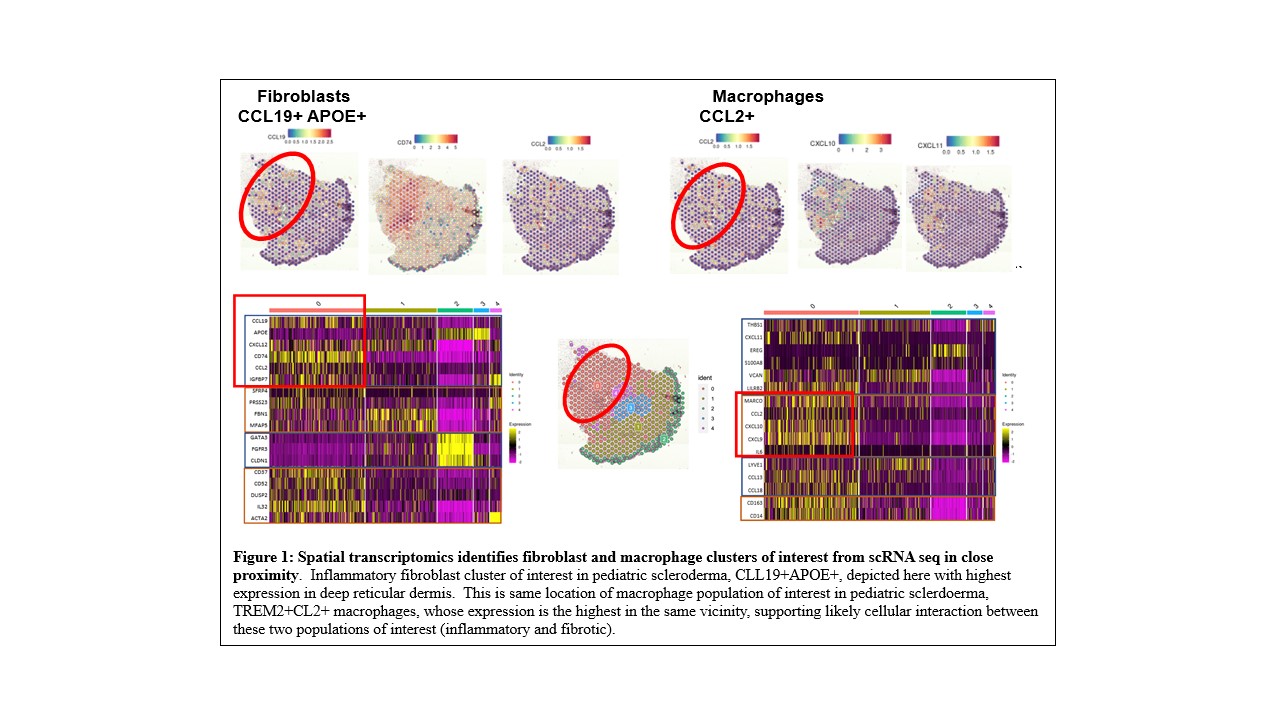
Results: We performed a pilot study on 4 skin FFPE samples, 3 from pediatric LS and 1 from pediatric SSc patients using the Visium 10X CytAssist for FFPE platform. We obtained an average of a total number of 566 spots under tissue section (4 mm x 4mm) with mean reads per spot 257,119, with 96% reads mapped to probe set, with 2,896 median genes detected per spot, and a total number of 18,029 genes detected. Transcriptomic expression using either marker genes or unbiased clustering was able to distinguish epidermis, dermis, and inflammatory regions. We also investigated some of the cell subsets and upregulated genes in those clusters from our scRNA seq data and observed the co-localization of an inflammatory fibroblast (CL19+APOE+) and a macrophage population (CCL2+TREM2+) that highly expresses IL-β in pediatric scleroderma together in the deep reticular dermis (Figure 1).

Conclusion: Using an innovative bioinformatic approach coupling scRNA seq and ST from paired tissue samples, we are able to identify dysregulated genes of interest, cells expressing these genes and their location within the skin and surrounding cells. To our knowledge, this would be the first study to employ spatial transcriptomics in localized scleroderma and systemic sclerosis skin.

Supporting image 1

Figure 1: Spatial transcriptomics identifies fibroblast and macrophage clusters of interest from scRNA seq in close proximity. Inflammatory fibroblast cluster of interest in pediatric scleroderma, CLL19+APOE+, depicted here with highest expression in deep reticular dermis. This is same location of macrophage population of interest in pediatric scleroderma, TREM2+CL2+ macrophages, whose expression is the highest in the same vicinity, supporting likely cellular interaction between these two populations of interest (inflammatory and fibrotic).


Disclosures: K. Torok: None; Y. Lai: None; Z. Xu: None; A. Sanyal: None; G. Werner: None; T. Hutchins: None; C. Cheng: None; W. Chen: None.

To cite this abstract in AMA style:

Torok K, Lai Y, Xu Z, Sanyal A, Werner G, Hutchins T, Cheng C, Chen W. Spatial Transcriptional Alterations in the Cellular Landscape of Pediatric Scleroderma Skin [abstract]. Arthritis Rheumatol. 2023; 75 (suppl 9). https://acrabstracts.org/abstract/spatial-transcriptional-alterations-in-the-cellular-landscape-of-pediatric-scleroderma-skin/. Accessed .
  • Tweet
  • Click to email a link to a friend (Opens in new window) Email
  • Click to print (Opens in new window) Print

« Back to ACR Convergence 2023

ACR Meeting Abstracts - https://acrabstracts.org/abstract/spatial-transcriptional-alterations-in-the-cellular-landscape-of-pediatric-scleroderma-skin/

Advanced Search

Your Favorites

You can save and print a list of your favorite abstracts during your browser session by clicking the “Favorite” button at the bottom of any abstract. View your favorites »

All abstracts accepted to ACR Convergence are under media embargo once the ACR has notified presenters of their abstract’s acceptance. They may be presented at other meetings or published as manuscripts after this time but should not be discussed in non-scholarly venues or outlets. The following embargo policies are strictly enforced by the ACR.

Accepted abstracts are made available to the public online in advance of the meeting and are published in a special online supplement of our scientific journal, Arthritis & Rheumatology. Information contained in those abstracts may not be released until the abstracts appear online. In an exception to the media embargo, academic institutions, private organizations, and companies with products whose value may be influenced by information contained in an abstract may issue a press release to coincide with the availability of an ACR abstract on the ACR website. However, the ACR continues to require that information that goes beyond that contained in the abstract (e.g., discussion of the abstract done as part of editorial news coverage) is under media embargo until 10:00 AM ET on November 14, 2024. Journalists with access to embargoed information cannot release articles or editorial news coverage before this time. Editorial news coverage is considered original articles/videos developed by employed journalists to report facts, commentary, and subject matter expert quotes in a narrative form using a variety of sources (e.g., research, announcements, press releases, events, etc.).

Violation of this policy may result in the abstract being withdrawn from the meeting and other measures deemed appropriate. Authors are responsible for notifying colleagues, institutions, communications firms, and all other stakeholders related to the development or promotion of the abstract about this policy. If you have questions about the ACR abstract embargo policy, please contact ACR abstracts staff at [email protected].

Wiley

  • Online Journal
  • Privacy Policy
  • Permissions Policies
  • Cookie Preferences

© Copyright 2025 American College of Rheumatology