Session Information
Session Type: Abstract Submissions (ACR)
Background/Purpose: Plantar fasciitis and Achilles tendonitis are commonly encountered in a rheumatologic practice due to either degenerative (DG) or systemic inflammatory conditions (SYS). While sonographic findings in both DG and SYS have been described in numerous studies, no study has systematically compared these two causes of heel pain. The aim of our study is to determine whether sonographic findings in the Achilles tendon and plantar fascia can be used to differentiate DG from SYS states.
Methods: Patients over the age of 18 with pain at the Achilles tendon or plantar fascia presenting to the Podiatry, Dermatology or Rheumatology clinics were enrolled. Exclusion criteria: previous heel trauma, surgery or recent corticosteroid heel injections. Medical chart review, a focused history, and a comprehensive musculoskeletal physical examination determined patient categorization as DG, SYS, or undetermined cause of heel pain. Patients’ Achilles tendons and plantar fascia were imaged with a GE Logiq e ultrasound and 12MHz linear array transducer. Doppler settings: PRF 0.8, low wall filter and Doppler gain set to maximize signal with minimal artifact. We evaluated tendon thickness, retro-calcaneal bursal size, cortical erosions, and Doppler signal at the enthesis and at the cortical margin. Continuous variable were compared between the degenerative and inflammatory groups using t tests, and proportions were compared using Chi squared or Fischer’s exact tests.
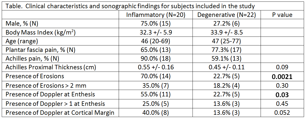
Results: Interim analysis of the first 46 patients recruited includes 20 in the SYS group and 22 in the DG group (4 undetermined). SYS group consists of 15 patients with psoriatic arthritis, 4 with non-psoriatic spondyloarthritis, and 1 with rheumatoid arthritis. Male:female ratio was 15:5 in the SYS group, and 6:16 in the DG group. While there were no significant differences in tendon thickness between the groups (Table), both the presence of erosions (SYS 70% vs. DG 23%, p=0.002), and the presence of Doppler at the enthesis (SYS 55%, vs. DG23%, p=0.03) were significantly more common in the SYS group (Table). Surprisingly, erosion size and degree of Doppler signal did not help distinguish the groups (Table). Doppler signal at the cortical margin was not more specific to the inflammatory group than enthesis Doppler overall (Table).
Conclusion: While there were no significant differences in tendon thickness, degenerative causes of heel enthesitis were less likely to show Doppler signal and erosive changes than systemic inflammatory enthesitis. However, erosions were seen in some heel enthesitis not associated with systemic inflammatory disease.
Disclosure:
P. Hook,
None;
D. Vradii,
None;
M. Dubreuil,
None;
H. Pham,
None;
E. Y. Kissin,
SonoSite Inc,
9.
« Back to 2014 ACR/ARHP Annual Meeting
ACR Meeting Abstracts - https://acrabstracts.org/abstract/sonographic-differentiation-of-heel-pain-focal-degenerative-versus-systemic-inflammatory-enthesitis/

