Session Information
Session Type: ACR Poster Session B
Session Time: 9:00AM-11:00AM
Background/Purpose:
The use of ultrasound in the assessment of muscle conditions has grown over the years. Various myopathies have shown an increase in echo intensity compared to healthy muscle1. The objective of this study was to compare muscles affected by myositis with normal controls on the basis of echo intensity, and to assess for differences in subjective appearance among particular forms of myositis.
Methods:
Sixty-two subjects were included in this study, consisting of 9 patient with polymyositis (PM), 7 with dermatomyositis (DM), 18 with inclusion body myositis (IBM), and 28 healthy controls. All patients met the ACR classification criteria for their particular disease. Seven muscle groups were examined bilaterally using ultrasound, including the deltoids, biceps, flexor carpi radialis (FCR), flexor digitorum profundus (FDP), rectus femoris, tibialis anterior and gastrocnemius muscles using a standardized protocol. Echo intensity was measured both subjectively using the Heckmatt scale (1-4)1 and quantitatively by mean gray-scale analysis using the program ITK-SNAP. Other parameters measured were duration of symptoms, muscle strength and muscle enzyme levels.
Results:
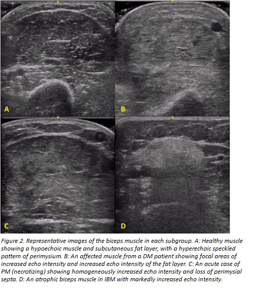
Echo intensity was increased in patients with myositis across all muscle groups studied when compared with normal muscles. Although numbers per subgroup were small, some visual patterns could be noted. Subcutaneous edema and increased echo intensity of fat could be seen in active DM. Focal areas of increased echogenicity were also noted within involved muscles, and other stigmata of DM such as calcinosis could be seen. PM, made up mostly of necrotizing myopathies, usually showed a more homogenous distribution of increased echo intensity in involved muscles and preserved fat echo intensity. IBM showed the most severely increased echo intensity and atrophy of the involved muscles.

Conclusion:
In the inflammatory myopathies, a higher echo intensity is seen in affected muscles. Additionally, disease
specific characteristics may be seen on visual inspection and can make ultrasound a useful tool in the
evaluation of muscle involvement in myositis.
1. Heckmatt et al. Detection of pathological change in dystrophic muscle with B-scan ultrasound imaging. Lancet 1, 1389–1390 (1980).
To cite this abstract in AMA style:
Leeuwenberg K, Christopher-Stine L, Bingham III CO, Paik JJ, Tiniakou E, Billings S, Burlina P, Albayda J. Sonographic Appearance of Inflammatory Myopathies: Increased Muscle Echointensity and Qualitative Changes [abstract]. Arthritis Rheumatol. 2018; 70 (suppl 9). https://acrabstracts.org/abstract/sonographic-appearance-of-inflammatory-myopathies-increased-muscle-echointensity-and-qualitative-changes/. Accessed .« Back to 2018 ACR/ARHP Annual Meeting
ACR Meeting Abstracts - https://acrabstracts.org/abstract/sonographic-appearance-of-inflammatory-myopathies-increased-muscle-echointensity-and-qualitative-changes/