ACR Meeting Abstracts

ACR Meeting Abstracts

  • Meetings
    • ACR Convergence 2024
    • ACR Convergence 2023
    • 2023 ACR/ARP PRSYM
    • ACR Convergence 2022
    • ACR Convergence 2021
    • ACR Convergence 2020
    • 2020 ACR/ARP PRSYM
    • 2019 ACR/ARP Annual Meeting
    • 2018-2009 Meetings
    • Download Abstracts
  • Keyword Index
  • Advanced Search
  • Your Favorites
    • Favorites
    • Login
    • View and print all favorites
    • Clear all your favorites
  • ACR Meetings

Abstract Number: 1173

Single-cell Multi-omic Analysis of a 3D Skin-Like Tissue Model Provides Insights into Molecular and Cellular Drivers of Systemic Sclerosis

Tamar Abel1, Noelle Kosarek2, Rezvan Parvizi3, Helen Jarnagin1, Mengqi Huang4, Avi Smith5, Michael Mariani1, Dillon Popovich6, Heetaek Yang7, Tammara Wood8, Jonathan Garlick9, Patricia Pioli7 and Michael Whitfield10, 1Dartmouth College, Lebanon, NH, 2Dartmouth Geisel School of Medicine, Lebanon, NH, 3Dartmouth, Lebanon, NH, 4University of Pittsburgh, Pittsburgh, PN, 5Tufts University, Boston, MA, 6Dartmouth College, West Lebanon, NH, 7Geisel School of Medicine at Dartmouth, Lebanon, NH, 8Dartmouth College, Hanover, NH, 9Tufts University School of Dental Medicine, Boston, MA, 10Geisel School of Medicine, Lebanon, NH

Meeting: ACR Convergence 2022

Keywords: Epigenetics, Fibroblasts, Other, Gene Expression, Systemic sclerosis, Tissue Engineering

  • Tweet
  • Click to email a link to a friend (Opens in new window) Email
  • Click to print (Opens in new window) Print
Session Information

Date: Sunday, November 13, 2022

Title: Systemic Sclerosis and Related Disorders – Basic Science Poster

Session Type: Poster Session C

Session Time: 1:00PM-3:00PM

Background/Purpose: Systemic Sclerosis (SSc) currently lacks reliable in vitro models of skin fibrosis constructed from all human cells. We have developed a skin-like tissue model of systemic sclerosis (SSc) that recapitulates the increased tissue thickness, stiffness, and molecular phenotype of SSc. Recent studies have identified specific pathogenic fibroblast (FB) subsets in skin and other affected tissues. The goal of this study was to characterize the cellular heterogeneity observed in our 3D tissues and compare these data to in vivo studies.  We hypothesize that our 3D model will in part recapitulate the FB heterogeneity observed in human skin and provide a tool for further insight into the cell-specific mechanisms of fibrosis in SSc.

Methods: Self-assembled Skin-equivalent (saSE) 3D tissues were grown in vitro over a period of 5 weeks by seeding healthy control (HC) or SSc FBs and monocytes into transwells, feeding with autologous plasma, and then layering normal human keratinocytes (NHKs) to form an epidermis. Following collagenase digestion of tissues, multi-omic scATAC-seq and RNA-seq data were generated using 10x protocols for a total of 3 biological replicates for both HC and SSc tissues. Statistical analyses of the single-cell dataset were performed in R using the Seurat and Signac packages. Samples were integrated and the ClusTree package was used to select the appropriate clustering resolution. Cell type was determined using cell type-specific gene expression.

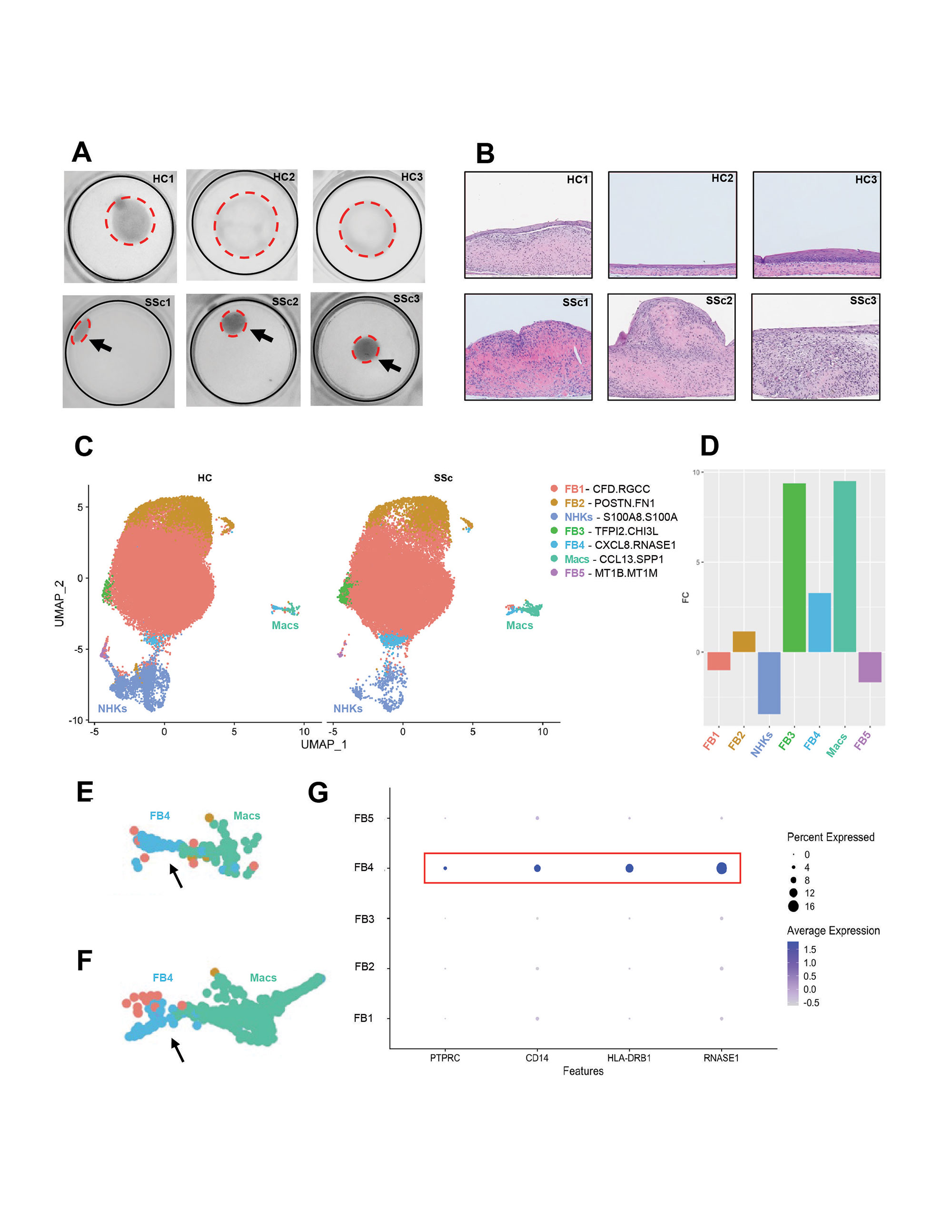
Results: Overhead images (Fig. 1A) and H&E histology (Fig. 1B) revealed a more contracted, thicker phenotype for SSc saSE skin-like tissues. Clustering resulted in identification of macrophage (Mac), NHK, and 4 major FB populations (Fig. 1C). Fold enrichment analysis showed a clear disease-specific enrichment of both Macs and 2 FB subsets (FB3 and FB5) (Fig. 1D). Cluster FB3 was the most enriched and displayed increased expression of SFRP4 and accessibility of the EGR1 and JUNB binding motifs. It was characterized by pathways associated with extracellular matrix proteins, proliferation, angiogenesis, and the recruitment of immune cells. FB5 cluster was also significantly enriched and was clustered adjacent to both FB and Mac populations (Fig. 1E-F). This population expressed collagen but was also enriched for myeloid genes including CD45, HLA-DRB1, and the monocyte marker CD14 (Fig. 1G). Pathways include wound healing, MHC II binding, and activation of T-cells. This cluster was not observed in scRNA-seq data from 3D tissues lacking the addition of monocytes.

Conclusion:

We identified 2 FB populations upregulated in the SSc saSE tissues. The FB3 subset highly expresses SFRP4 and may recapitulate a population recently identified as enriched in SSc skin via single-cell analysis. Epigenetic data suggests that EGR1 and JUNB may play a critical role in maintaining this FB state. In addition, we identify enrichment of a novel FB subset, FB5, with markers of both myeloid and mesenchymal cells. Most importantly, we were able to characterize the FB heterogeneity of our 3D skin-like tissue model confirming that this model can approximate the cellular complexity observed in human skin and may serve as a suitable model for additional studies of pathogenic FB subsets in SSc. 

Supporting image 1

Figure 1. Analysis of 3D tissue cell clusters based on single-cell epigenomic and transcriptomic profiles reveals Systemic sclerosis (SSc)-specific fibroblast subset enrichment. A) Overhead images of 3D tissues in transwell insert. Three healthy control (HC) biological replicates (HC1, HC2, HC3) and three SSc biological replicates (SSc1, SSc2, SSc3). Black line indicates border of insert membrane and red dotted line indicates borders of 3D tissue. B) H&E histology of 3D tissues showing representative sections for each biological replicate. C) UMAP projection of cells clustered based on transcriptional data (n=6) and split by disease state. Normal human keratinocytes (NHKs) and Macrophages (Macs) are labeled in text matching the color of each respective cluster. All other clusters are fibroblasts as determined by cell-specific gene expression. Legend on the right includes cell cluster labels as well as the top two differentially expressed genes in that cluster as compared to all other clusters. D) Fold change graph for each cluster in SSc 3D tissues (n=3) as compared to HC tissues (n=3). Macs, FB3, and FB4 are significantly enriched in SSc tissues while most other clusters are slightly decreased. Close up view of adjoining FB4 and Mac clusters in E) HC tissues and F) SSc tissues with arrows indicating location of FB4 cluster. G) Expression of myeloid genes compared across fibroblast subsets shows increased expression in FB4 population (highlighted by red box).


Disclosures: T. Abel, None; N. Kosarek, None; R. Parvizi, None; H. Jarnagin, None; M. Huang, None; A. Smith, None; M. Mariani, None; D. Popovich, None; H. Yang, None; T. Wood, None; J. Garlick, None; P. Pioli, Celdara Medical, LLC; M. Whitfield, Bristol-Myers Squibb(BMS), Celdara Medical LLC.

To cite this abstract in AMA style:

Abel T, Kosarek N, Parvizi R, Jarnagin H, Huang M, Smith A, Mariani M, Popovich D, Yang H, Wood T, Garlick J, Pioli P, Whitfield M. Single-cell Multi-omic Analysis of a 3D Skin-Like Tissue Model Provides Insights into Molecular and Cellular Drivers of Systemic Sclerosis [abstract]. Arthritis Rheumatol. 2022; 74 (suppl 9). https://acrabstracts.org/abstract/single-cell-multi-omic-analysis-of-a-3d-skin-like-tissue-model-provides-insights-into-molecular-and-cellular-drivers-of-systemic-sclerosis/. Accessed .
  • Tweet
  • Click to email a link to a friend (Opens in new window) Email
  • Click to print (Opens in new window) Print

« Back to ACR Convergence 2022

ACR Meeting Abstracts - https://acrabstracts.org/abstract/single-cell-multi-omic-analysis-of-a-3d-skin-like-tissue-model-provides-insights-into-molecular-and-cellular-drivers-of-systemic-sclerosis/

Advanced Search

Your Favorites

You can save and print a list of your favorite abstracts during your browser session by clicking the “Favorite” button at the bottom of any abstract. View your favorites »

All abstracts accepted to ACR Convergence are under media embargo once the ACR has notified presenters of their abstract’s acceptance. They may be presented at other meetings or published as manuscripts after this time but should not be discussed in non-scholarly venues or outlets. The following embargo policies are strictly enforced by the ACR.

Accepted abstracts are made available to the public online in advance of the meeting and are published in a special online supplement of our scientific journal, Arthritis & Rheumatology. Information contained in those abstracts may not be released until the abstracts appear online. In an exception to the media embargo, academic institutions, private organizations, and companies with products whose value may be influenced by information contained in an abstract may issue a press release to coincide with the availability of an ACR abstract on the ACR website. However, the ACR continues to require that information that goes beyond that contained in the abstract (e.g., discussion of the abstract done as part of editorial news coverage) is under media embargo until 10:00 AM ET on November 14, 2024. Journalists with access to embargoed information cannot release articles or editorial news coverage before this time. Editorial news coverage is considered original articles/videos developed by employed journalists to report facts, commentary, and subject matter expert quotes in a narrative form using a variety of sources (e.g., research, announcements, press releases, events, etc.).

Violation of this policy may result in the abstract being withdrawn from the meeting and other measures deemed appropriate. Authors are responsible for notifying colleagues, institutions, communications firms, and all other stakeholders related to the development or promotion of the abstract about this policy. If you have questions about the ACR abstract embargo policy, please contact ACR abstracts staff at [email protected].

Wiley

  • Online Journal
  • Privacy Policy
  • Permissions Policies
  • Cookie Preferences

© Copyright 2025 American College of Rheumatology