Session Information
Date: Sunday, October 21, 2018
Title: Systemic Lupus Erythematosus – Clinical Poster I: Clinical Manifestations and Comorbidity
Session Type: ACR Poster Session A
Session Time: 9:00AM-11:00AM
Background/Purpose: Recent studies suggest that autophagy defects contributes to systemic lupus erythematosus (SLE) pathogenesis. Prolactin (PRL) is associated with active SLE and stimulates the immune cells by binding to receptor (PRL-R). Prolactin decreases the suppressor function exerted by Treg cells favoring an inflammatory microenvironment in SLE. The objective is simultaneously identify the expression of autophagy in Treg lymphocytes and PRL-R in B lymphocytes of SLE patients.
Methods: We included 40 patients with SLE (ACR criteria) divided into two groups: Group 1: patients with remission SLE (SLEDAI <4), Group 2: patients with active SLE (SLEDAI > 4). The affected organs and the treatments received were obtained. As a control group, we included healthy individuals. A fasting peripheral blood sample was taken from all patients and controls. The cells were separated and labeled with specific antibodies: Treg cells: CD25+, FoxP3+, Atg14+ (autophagy marker), and B lymphocytes: CD19+, PRL-R+. Analysis was performed by flow cytometry. ANOVA test was used for statistical analysis.
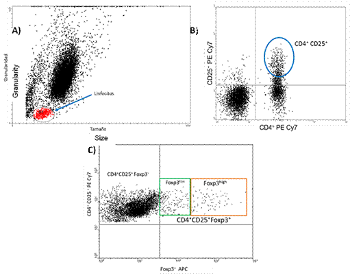
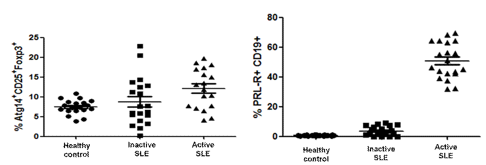
Results: We included 40 patients, divided in 20 inactive, 20 active SLE patients and 20 healthy controls. 80% of our population were female. Evolution time in inactive SLE patients was 8.72±1.16 years and in active SLE patients was 5±0.95 years. SLEDAI was higher in active SLE patients than inactive SLE patients. In both active and inactive SLE groups, kidney was the most affected major organ (55% and 25% respectively). Sixty percent of inactive SLE patients and 35% of active patients had Chloroquine. The expression of autophagy, Treg cells Prolactin receptors and B-lymphocytes are shown in Figures 1 and 2.
Conclusion: The increase of autophagy in Treg and PRL-R in B lymphocytes participate in active lupus glomerulonephritis. Autophagy and PRL-R may be new therapeutic targets in SLE.This results suggest that may be a connection between autophagy, PRL-R, Treg and B cells if any.
Fig.1: A) Dot plot in logarithmic scale of peripheral blood mononuclear cells (PBMC) of a SLE patient. B) Dot plot of the percentage of CD4 + CD25 + expression for Treg lymphocytes. C) Dot plot of the percentage of CD4 + CD25 + expression for Treg lymphocytes
Fig. 2: Left: autophagy (%) in Treg cells in patients and controls: A: Healthy control vs. Inactive SLE: 7.44 vs 8.74 p= >0.9999 B: Healthy control vs. Active SLE: 7.44 vs 12.11 p= <0.05. Right: prolactin Receptor in B lymphocytes, in Active and Inactive patients and healthy controls: A: Healthy Controls vs Inactive SLE: 0.62 vs 3.56 p= 0.7463. B: Heatlhy Controls vs Active SLE: 0.62 vs 50.79 p= <0.0001
To cite this abstract in AMA style:
Jara-Quezada LJ, Zurita E, Durán A, Sanchez A, Bustamante R, Medina G, Saavedra MA, Cruz-Dominguez MP, Vera-Lastra O, Jiménez-Arellano MP, Martínez-Bencomo MA, Rodriguez A. Simultaneous Identification of Two Biomarkers in ACTIVE LUPUS Nephritis: Autophagy in Treg CELLS and Prolactin Receptors in B Lymphocytes [abstract]. Arthritis Rheumatol. 2018; 70 (suppl 9). https://acrabstracts.org/abstract/simultaneous-identification-of-two-biomarkers-in-active-lupus-nephritis-autophagy-in-treg-cells-and-prolactin-receptors-in-b-lymphocytes/. Accessed .« Back to 2018 ACR/ARHP Annual Meeting
ACR Meeting Abstracts - https://acrabstracts.org/abstract/simultaneous-identification-of-two-biomarkers-in-active-lupus-nephritis-autophagy-in-treg-cells-and-prolactin-receptors-in-b-lymphocytes/


