Session Information
Session Type: ACR Poster Session A
Session Time: 9:00AM-11:00AM
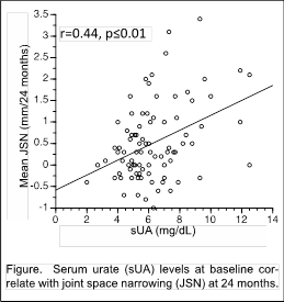
Background/Purpose: Osteoarthritis (OA) etiopathogenesis includes an inflammatory component. Published reports indicate that synovial fluid urate levels, even in patients without gout, associate with OA prevalence/severity. Whether serum urate (sUA), the precursor for gout and a biomarker for cardiovascular and kidney disease, may serve as a biomarker to convey or predict OA risk is not known. We investigated whether sUA levels associate with knee OA radiographic severity and contrast MRI-measured quantitative synovial volume (SV), and whether sUA levels predict radiographic progression, in a gout-free knee OA cohort.
Methods: We assessed sUA in 88 gout-free subjects who completed a 24-month prospective, natural history knee OA study. Subjects had symptomatic medial knee OA, met ACR knee OA criteria and had BMI <33 at study entry. sUA was measured (enzyme-colorimetry) in serum frozen and banked at baseline. At baseline and 24 months, patients underwent standardized weight-bearing fixed-flexion posteroanterior knee radiographs (SynaFlexer™). Twenty-seven subjects additionally had a dynamic gadolinium-enhanced 3.0T knee MRI that was read for quantitative synovial volume (SV). A musculoskeletal radiologist, blinded to subject data, determined joint space width (JSW) and Kellgren-Lawrence (KL) grades at each time point. Joint space narrowing (JSN) was determined as JSW change from baseline to 24 months. Pearson’s correlations, student’s t-tests, one-way ANOVA with post hoc Tukey-Kramer tests, ROC and AUC curves were used in statistical analyses, as appropriate.
Results: sUA correlated with JSN in both univariate (r=0.40, p≤0.01) and multivariate analyses (adjusting for age, gender and BMI, r=0.28, p=0.010). There was a significant difference in mean JSN after dichotomization of sUA at 6.8mg/dL, the solubility point for serum urate, even after adjustment for age, gender and BMI (JSN [±SEM] of 0.90mm±0.20mm for sUA≥6.8; JSN [±SEM] of 0.31mm±0.09mm for sUA<6.8, p<0.01). Baseline sUA distinguished progressors (JSN>0.2mm), and fast progressors (JSN>0.5mm), from non-progressors (JSN≤0.0mm) in multivariate analyses (area under the receiver operating characteristic curve [AUC] 0.626, p=0.027; AUC 0.620, p=0.045, respectively). sUA also correlated with SV (r=0.44, p=0.0040), a possible marker of JSN, though this correlation did not persist after controlling for age, gender and BMI (r=0.13, p=0.562).
Conclusion: In non-gout patients with knee OA, sUA levels predict JSN and may serve as a biomarker for OA progression. 
To cite this abstract in AMA style:
Oshinsky C, Attur M, Ma S, Zhou H, Zheng F, Chen M, Patel J, Samuels J, Pike V, Regatte R, Bencardino J, Rybak L, Abramson SB, Pillinger MH, Krasnokutsky Samuels S. Serum Urate Levels Predict Joint Space Narrowing in Non-Gout Patients with Medial Knee Osteoarthritis [abstract]. Arthritis Rheumatol. 2016; 68 (suppl 10). https://acrabstracts.org/abstract/serum-urate-levels-predict-joint-space-narrowing-in-non-gout-patients-with-medial-knee-osteoarthritis/. Accessed .« Back to 2016 ACR/ARHP Annual Meeting
ACR Meeting Abstracts - https://acrabstracts.org/abstract/serum-urate-levels-predict-joint-space-narrowing-in-non-gout-patients-with-medial-knee-osteoarthritis/
