Session Information
Date: Sunday, November 17, 2024
Title: Systemic Sclerosis & Related Disorders – Basic Science Poster I
Session Type: Poster Session B
Session Time: 10:30AM-12:30PM
Background/Purpose: Systemic sclerosis (SSc) is a diffuse connective tissue disease associated with multi-system and multi-organ tissue damage. The transformation of different precursor cells to myofibroblasts is one of the key factors in the pathogenesis of SSc-PF. In this study, we intend to further validate the existence of lung basal cell transformation to myofibroblasts in SSc-PF, to clarify the phenotypic transformation and molecular mechanism of TGF-β1-induced lung basal cell transformation, to verify the role of lung basal cells in the pathogenesis of SSc-PF through in vivo experiments, and to search for possible molecular targets.
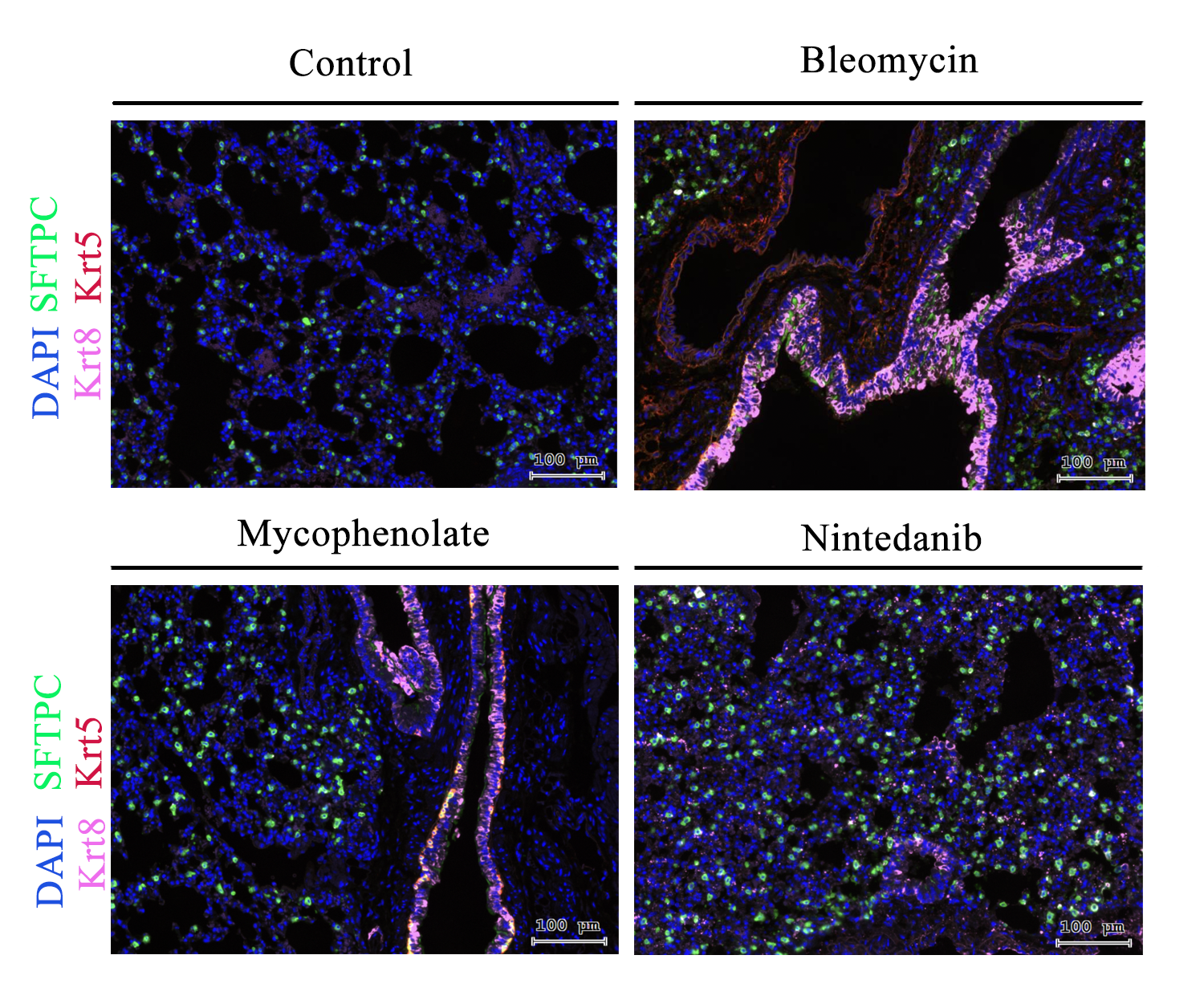
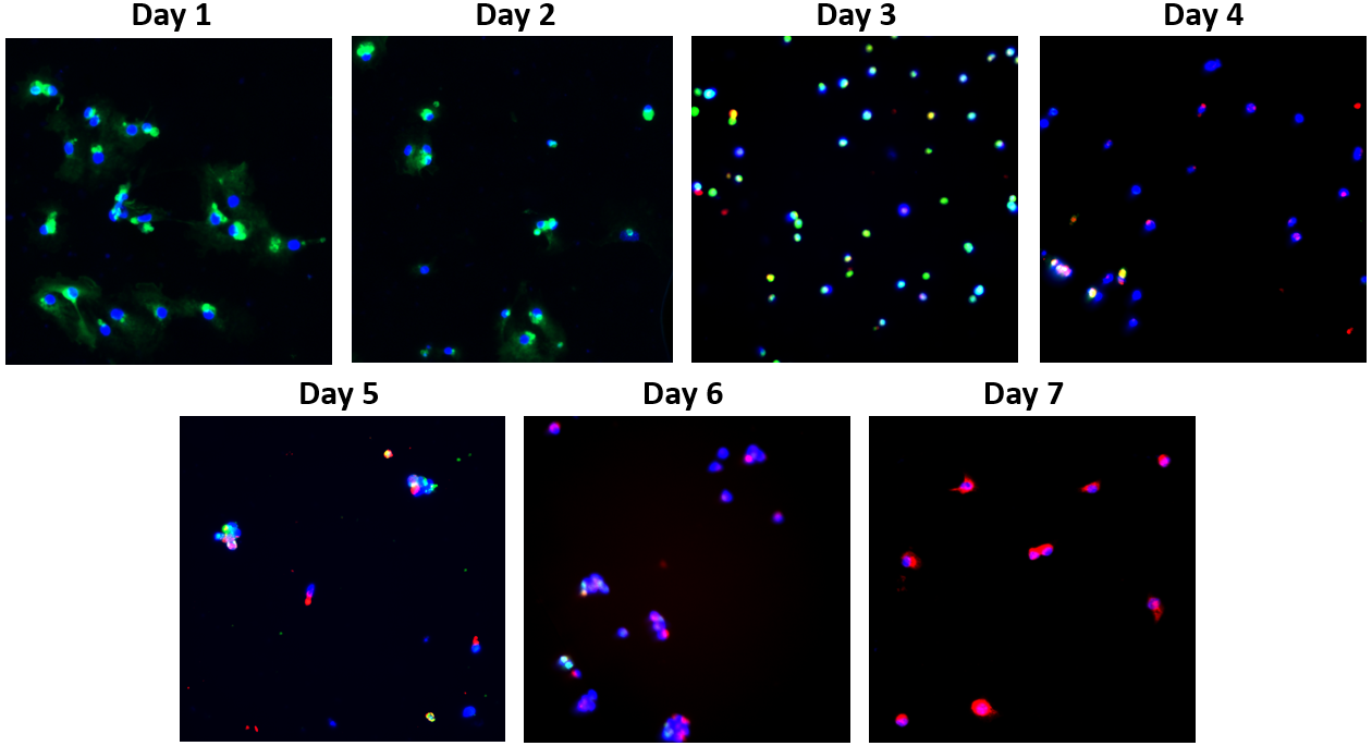
Methods: Single-cell sequencing analysis was performed with our ssc lung transplantation samples and public database data to identify the presence of intermediate state cells and screen for possible pathways; a model of systemic sclerosis was established by subcutaneous injection of bleomycin method in BALB/C mice, and the lungs were stained with type II alveolar epithelial cells (SFTPC), lung basaloidal cells (Krt5), and lung intermediate state cell markers (Krt8), and DAPI for Immunofluorescence staining was performed to observe the differences in the proportion and number of intermediate state cells in different drug-administered groups; TGF-β1 (10 ng/mL) was used for 7 days to observe the changes in fluorescence intensity over time.
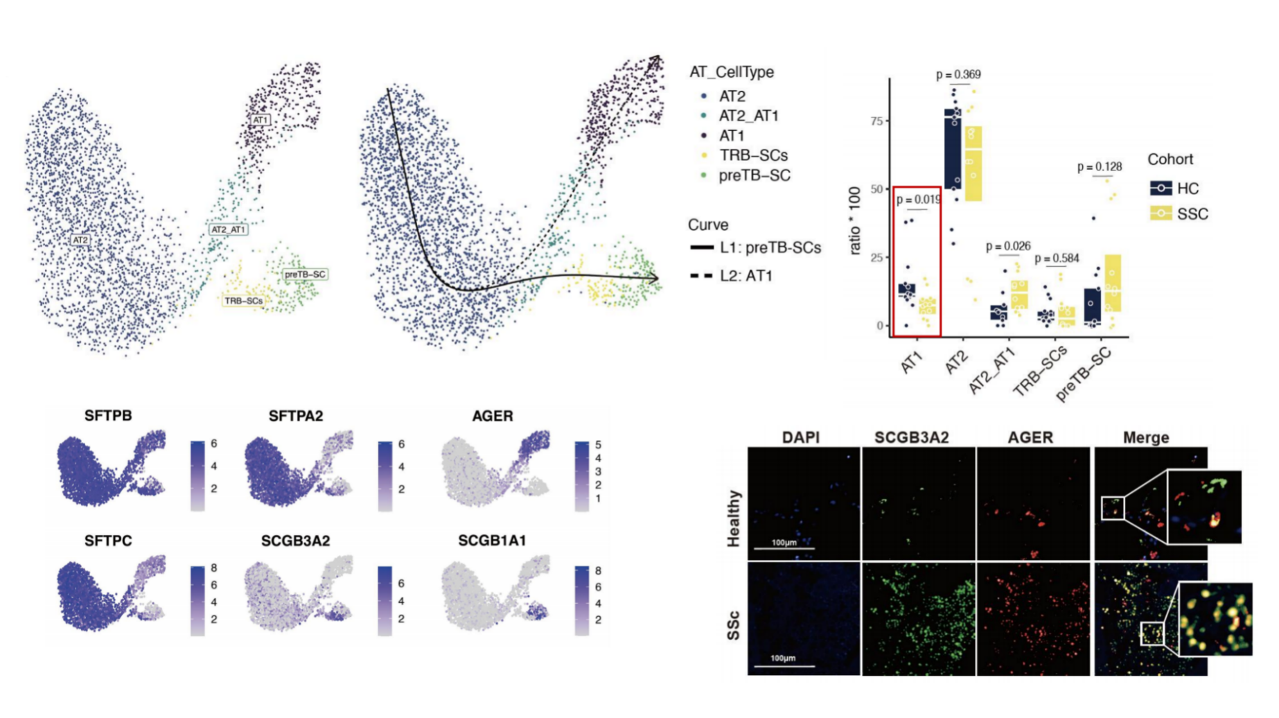
Results: Previous studies found that alveolar type II epithelial cells (AT2) in the process of differentiation to alveolar AT1 in the presence of external stimuli to the presence of intermediate state that is Krt8 + Krt5-SFTPC + orlowAT, presumably the number and functional changes associated with the pathological process of fibrosis of the SSc-PF lungs, and regulated by the Wnt3a-FZD5 pathway (Fig. 1); immunofluorescence results reality, the and compared with the lung tissues of the control (saline intervention) group, there was a significant increase in mouse lung basal cells after bleomycin action and co-localisation with the type II alveolar epithelial cell fraction, and a significant decrease in the lung basal cells after the treatment group (nidanib or mottled mescaline ester intervention) (Fig. 2). In addition, fluorescent staining with type II alveolar epithelial cell marker (SFTPC) and lung basal cell marker (Krt5) as well as DAPI was performed after 7 days of intervention with TGF-β1 (10 ng/mL), during which a gradual weakening of the type II alveolar epithelial cell marker (SFTPC) and gradual enhancement of the lung basal cell marker (Krt5) was observed, with the majority of type II alveolar epithelial cells being transformed into lung basal cells (Fig. 2) by day 7. transformed into lung basal cells (Figure 3).
Conclusion: The number and proportion of intermediate state cells during the transformation of lung basal cells to fibroblasts plays an important role in the pathogenesis of systemic sclerosis and may regulate the cellular transformation process through the Wnt3a-FZD5 pathway.
To cite this abstract in AMA style:
Yu H, Xue J. Role and Mechanism of the Wnt3a-FZD5 Pathway in Regulating the Transformed Intermediate State of Alveolar Epithelial Cells Involved in Pulmonary Fibrosis in Systemic Sclerosis [abstract]. Arthritis Rheumatol. 2024; 76 (suppl 9). https://acrabstracts.org/abstract/role-and-mechanism-of-the-wnt3a-fzd5-pathway-in-regulating-the-transformed-intermediate-state-of-alveolar-epithelial-cells-involved-in-pulmonary-fibrosis-in-systemic-sclerosis/. Accessed .« Back to ACR Convergence 2024
ACR Meeting Abstracts - https://acrabstracts.org/abstract/role-and-mechanism-of-the-wnt3a-fzd5-pathway-in-regulating-the-transformed-intermediate-state-of-alveolar-epithelial-cells-involved-in-pulmonary-fibrosis-in-systemic-sclerosis/


