ACR Meeting Abstracts

ACR Meeting Abstracts

  • Meeting Abstracts
    • All Meetings
    • PRSYM 2026
    • ACR Convergence 2025
    • Download Abstract Supplements
  • Keyword Index
  • Search
  • My Favorites
    • View and print all favorites
    • Clear all favorites

Abstract Number: 1501

Retrospective Cohort Study Identifying Pulmonary Complications in a Cohort of Patients with Systemic Lupus Erythematosus

Jessica Johnson1, Chao Zhang2 and Emily Littlejohn3, 1Cleveland Clinic Lerner College of Medicine, Cleveland, OH, 2Cleveland Clinic, Cleveland Heights, OH, 3Cleveland Clinic, Cleveland, OH

Meeting: ACR Convergence 2024

Keywords: Clinical practice guidelines, pulmonary, risk factors, Systemic lupus erythematosus (SLE)

  • Tweet
  • Email a link to a friend (Opens in new window) Email
  • Print (Opens in new window) Print
Session Information

Date: Sunday, November 17, 2024

Title: SLE – Diagnosis, Manifestations, & Outcomes Poster II

Session Type: Poster Session B

Session Time: 10:30AM-12:30PM

Background/Purpose: Systemic lupus erythematosus (SLE) is a systemic autoimmune disease with multi organ involvement. One of the most common manifestations is pulmonary disease with a reported prevalence between 5 – 90%. Given this wide range of prevalence, there is a need to more closely define types of pulmonary disease in SLE and associated risk factors. We sought to characterize the presentation of pulmonary manifestations in an established SLE cohort using electronic health record data.

Methods: A retrospective chart review was performed using The Cleveland Clinic Health System electronic medical record data and the Cleveland Clinic Lupus Cohort (CCLC) data. All patients were >18 years of age and had confirmed SLE by a Cleveland clinic foundation rheumatologist using the 2012 SLICC or 2019 ACR/EULAR classification criteria. The cohort included 313 patients, 220 of whom had imaging studies completed. Generalized estimating equations (GEE) were utilized to analyze the data, accounting for its repeated measured nature.

Results: The average age in our cohort was 42.5 years, 86.7% identified as female, 60.5% identified as white, 37.3% as Black, and 1.82% as Asian (Table 1). The most common findings on CT and x-ray were increased lung density (24%, 12%) and atelectasis (18%, 10%). The most common disease found on CT was pleural effusion (24%) or mediastinal/axillary lymphadenopathy (16%). Our GEE analysis showed no association between demographics and CT findings; however, patients who had smoked (current or former), had higher odds (OR 2.28, 95% CI: 1.05 – 4.94, p = 0.04) of radiologist reported disease on their imaging compared with patients who never smoked, even after adjusting for other covariates (Table 2). Additionally, the GEE results for disease on x-ray, as reported by the radiologist, showed that age, gender, smoking status, and x-ray time were significantly different in those who had a disease reported on their radiological report compared with those who did not (Table 3).

Conclusion: With the limited literature commenting on pulmonary involvement in SLE, our results show that certain factors, namely smoking, older age, and male sex should prompt clinicians to have a higher suspicion for lung disease in SLE patients. While our study is limited by the retrospective nature, lack of consistent SLEDAI-2K scores, and medication information within a date considered acceptable for inclusion in our analysis, our results highlight important characteristics for predicting pulmonary involvement in SLE. It is also important to note that the pathologies described herein cannot be directly attributed to SLE and could have secondary causes such as infection or other organ system failure. Further characterization in larger cohorts should be conducted to explore these possible associations. Until then, awareness of these potential risks is important for clinicians caring for SLE patients to optimize their care and follow up.
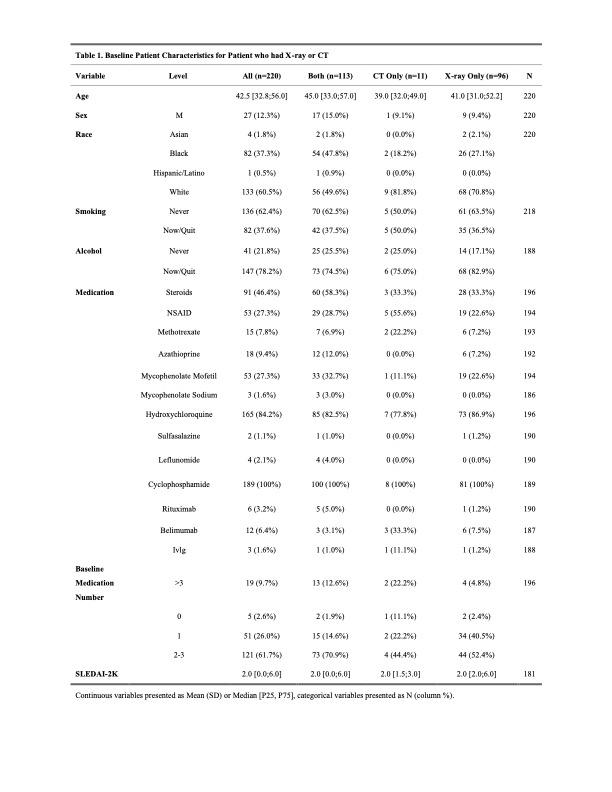
Supporting image 1

Table 1. Baseline Patient Characteristics for the Imaging Dataset. Baseline demographic information for all patients in the registry dataset who had imaging completed are reported. 220 of 313 had either CT, X-ray, or both completed prior to the collection date. Some patients did not have complete baseline demographic information.

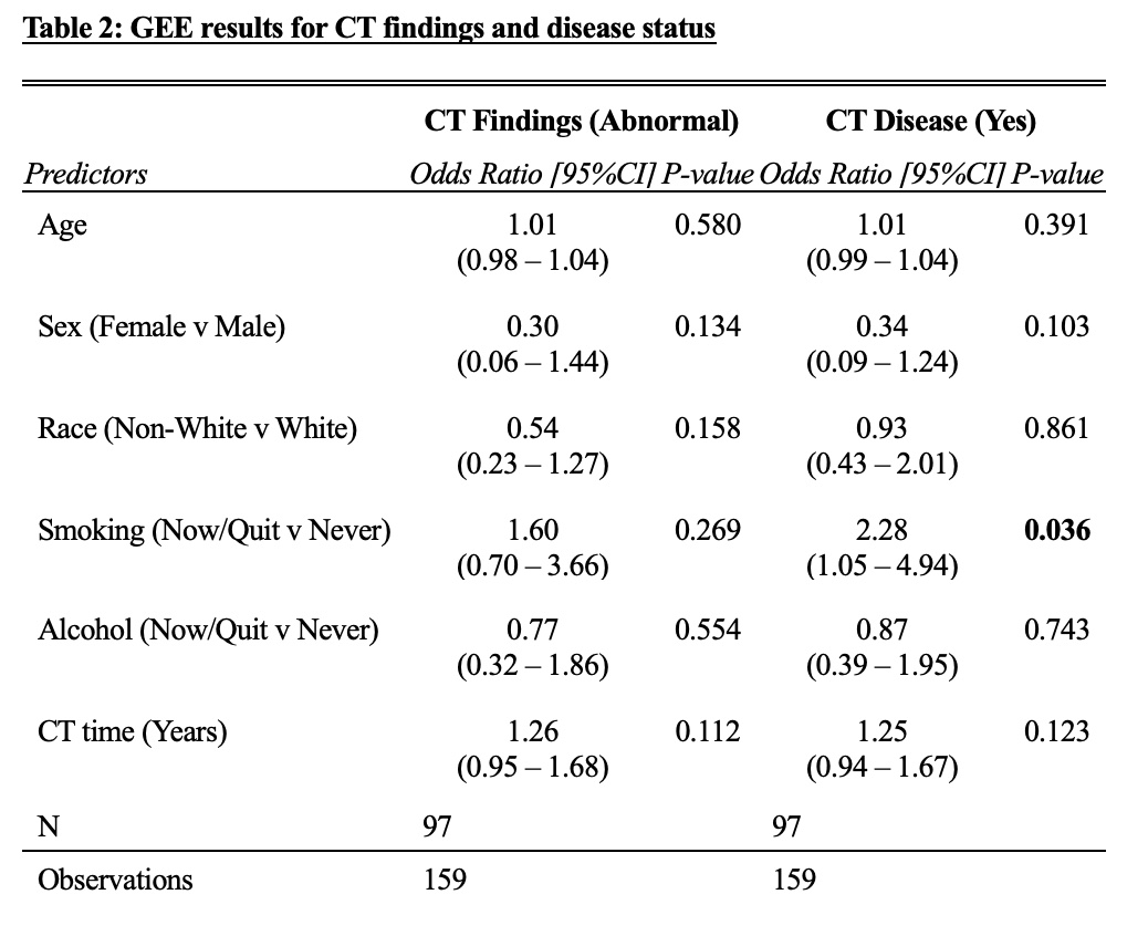
Supporting image 2

Table 2. GEE results for CT findings and disease status. Within the GEE model for CT imaging data, 97 patients had complete information and were included in the analysis. The outcome of interest was CT finding (abnormal vs normal) and evidence of CT disease (yes vs no). The results showed no association between demographics and CT findings; however, patients who had smoked (current or former), had higher odds (OR 2.28, 95% CI: 1.05 – 4.94, p = 0.04) of radiologist reported disease on their imaging compared with patients who never smoked, even after adjusting for other covariates.

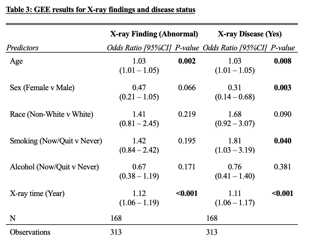
Supporting image 3

Table 3. GEE results for x-ray findings and disease status. 168 patients and their corresponding x-ray results were included in the analysis. The outcome of interest is x-ray finding (abnormal versus normal) and x-ray disease (yes versus no). The results for x-ray disease, as reported by the radiologist, showed that age, gender, smoking status, and x-ray time were significantly different in those who had a disease reported on their radiological report versus those who did not. For age, each additional year of age was associated with a higher odd (OR: 1.03, 95% CI: 1.01 – 1.05, p = 0.008) of disease, after adjusting for other covariates. Females were less likely to have disease compared to males (OR: 0.31, 95% CI: 0.14 – 0.68, p = 0.003). Patients who ever smoked or currently smoked had higher odds (OR: 1.81, 95% CI: 1.03 – 3.19, p = 0.04) of disease compared with never smokers. Each additional year of follow-up time was associated with an 11% (OR: 1.11, 95% CI: 1.06 – 1.17, p < 0.001) increase in odds of having a disease reported on the radiology report, after adjusting for other covariates.


Disclosures: J. Johnson: None; C. Zhang: None; E. Littlejohn: Aurinia Pharmaceuticals, 6, Bristol-Myers Squibb(BMS), 5, GlaxoSmithKlein(GSK), 2, Synthekine, 2, 5.

To cite this abstract in AMA style:

Johnson J, Zhang C, Littlejohn E. Retrospective Cohort Study Identifying Pulmonary Complications in a Cohort of Patients with Systemic Lupus Erythematosus [abstract]. Arthritis Rheumatol. 2024; 76 (suppl 9). https://acrabstracts.org/abstract/retrospective-cohort-study-identifying-pulmonary-complications-in-a-cohort-of-patients-with-systemic-lupus-erythematosus/. Accessed .
  • Tweet
  • Email a link to a friend (Opens in new window) Email
  • Print (Opens in new window) Print

« Back to ACR Convergence 2024

ACR Meeting Abstracts - https://acrabstracts.org/abstract/retrospective-cohort-study-identifying-pulmonary-complications-in-a-cohort-of-patients-with-systemic-lupus-erythematosus/

Advanced Search

My Favorites

Save and print abstracts during your browser session by clicking the “Favorite” button at the bottom of any abstract (must have cookies enabled in browser). See saved favorites.

Abstract Policies

  • ACR Convergence Abstract Embargo Policies
  • ACR Convergence Abstract Permissions & Reprints
  • PRSYM Abstract Policies

ACR Convergence. Where Rheumatology Meets

ACR Convergence 2026

Join us November 6-11 in Orlando, Florida.
See Registration Information

  • Contact ACR
  • Privacy Policy
  • ACR Policies
  • Cookie Preferences

© Copyright 2026 American College of Rheumatology