Session Information
Session Type: ACR Poster Session C
Session Time: 9:00AM-11:00AM
Background/Purpose: Gout duration and serum urate (SU) levels are thought to influence development of tophi and chronic inflammatory gouty arthropathy, but the extent to which either one or both contribute to their development is not clear. We evaluated the relation of SU and of gout duration to the presence of tophus, urate deposition, and inflammation in a large cohort of crystal-proven gout subjects.
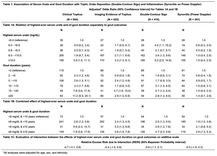
Methods: We used data from the 509 crystal-proven gout subjects in the Study for Updated Gout Classification Criteria (SUGAR) cohort to evaluate the effect of highest-ever recorded SU and gout duration (years since 1st flare) separately, as well as in combination to examine their joint effects on tophi, urate deposition, and inflammation. Subjects were enrolled irrespective of flare status. Radiographs of the hands and feet, and ultrasound (US) of a clinically affected joint were obtained. Tophi were evaluated as clinically evident tophi, as well as imaging evidence of tophus (based on X-ray or US). Urate deposition was determined based on US evidence of urate deposition (double-contour sign), and inflammation was assessed as power Doppler signal on US indicative of synovitis. We assessed the relation of highest SU, gout duration, and their combination using logistic regression, adjusting for age, sex, and ethnicity. Relative Excess Risk due to Interaction (RERI) was assessed using a linear additive risk-ratio model.
Results: Study sample included ~87% males, ~66% Caucasians, mean age of 60 years. Highest SU ranged from 2.6-19.2 mg/dL, and gout duration from 0-54 yrs. Higher levels of SU and longer duration of gout were significantly associated with clinical tophus, imaging evidence of tophus and double-contour sign, but not with power Doppler signal (Table 1A). The combination of higher SU and longer gout duration had the strongest association with the 4 outcomes, followed by longer disease duration regardless of highest SU for tophi and urate deposition, while for synovitis effects were similar regardless of the combination of SU with gout duration compared with low SU and shorter gout duration (Table 1B). However, we did not find any evidence of an additive interaction (Table 1C).
Conclusion: Both gout duration and highest-ever recorded SU levels were each associated with clinical and imaging evidence of tophus and with US-evidence of urate deposition, but not clearly with evidence of synovitis (power Doppler). The combination of high SU and longer gout duration resulted in the highest risk of tophaceous disease, but there not appeared to be a potentiation of individual effects. These data highlight the importance of managing the hyperuricemia of gout to minimize the adverse clinical features of the disease.
To cite this abstract in AMA style:
Vargas-Santos AB, Jafarzadeh SR, Castelar-Pinheiro G, Dalbeth N, Taylor WJ, Fransen J, Jansen TL, Schumacher HR, Neogi T. Relation of Serum Urate and Gout Duration to Tophi, Urate Deposition, and Inflammation [abstract]. Arthritis Rheumatol. 2017; 69 (suppl 10). https://acrabstracts.org/abstract/relation-of-serum-urate-and-gout-duration-to-tophi-urate-deposition-and-inflammation/. Accessed .« Back to 2017 ACR/ARHP Annual Meeting
ACR Meeting Abstracts - https://acrabstracts.org/abstract/relation-of-serum-urate-and-gout-duration-to-tophi-urate-deposition-and-inflammation/

