ACR Meeting Abstracts

ACR Meeting Abstracts

  • Meetings
    • ACR Convergence 2024
    • ACR Convergence 2023
    • 2023 ACR/ARP PRSYM
    • ACR Convergence 2022
    • ACR Convergence 2021
    • ACR Convergence 2020
    • 2020 ACR/ARP PRSYM
    • 2019 ACR/ARP Annual Meeting
    • 2018-2009 Meetings
    • Download Abstracts
  • Keyword Index
  • Advanced Search
  • Your Favorites
    • Favorites
    • Login
    • View and print all favorites
    • Clear all your favorites
  • ACR Meetings

Abstract Number: 1804

Raman Spectroscopy Integrated with Polarized Light Microscopy for Diagnosis of Crystallopathies

Tom Niessink1, Matthijs Janssen2, Cees Otto1 and Tim Jansen3, 1Medical Cell Biophysics group, University of Twente, Enschede, Netherlands, 2Rijnstate Hospital, Bennekom, Netherlands, 3Department of Rheumatology, VieCuri Medical Centre, Venlo, Netherlands

Meeting: ACR Convergence 2022

Keywords: Crystal-induced arthritis, gout, pseudogout

  • Tweet
  • Click to email a link to a friend (Opens in new window) Email
  • Click to print (Opens in new window) Print
Session Information

Date: Monday, November 14, 2022

Title: Metabolic and Crystal Arthropathies – Basic and Clinical Science Poster

Session Type: Poster Session D

Session Time: 1:00PM-3:00PM

Background/Purpose: The current golden standard in diagnosing gout and calcium pyrophosphate deposition is polarized light microscopy (PLM). However, small crystal sizes, the presence of other crystal types, glass artefacts and cellular uptake of MSU can pose uncertainties in the clinical analysis of synovial fluids. There are multiple methods to improve synovial fluid analysis, of which Raman spectroscopy is the most promising. In our approach, we have integrated an ordinary polarized light microscope with a Raman spectroscope (iRPolM, Hybriscan), allowing for simultaneous use of both techniques. In this abstract, we demonstrate the added value of this technique for the first 50 patients, including 23 diagnosed with gout.

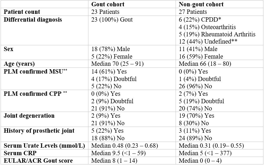
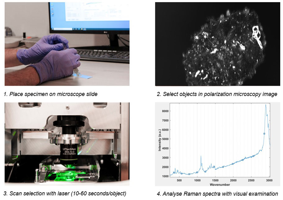
Methods: 50 patients were punctured for diagnostic purposes when presenting with swollen joints. Patient characteristics are given in figure 1. Samples were analyzed by an experienced rheumatologist with polarization microscopy, who also gave an indication of certainty in diagnosis. The iRPolM analysis followed this. A visual representation of this method can be found in figure 2. Retrieved Raman spectra were analyzed by a trained Raman spectroscopist for the presence of MSU, calcium pyrophosphate dihydrate (CPPD) or other (deposited) materials.

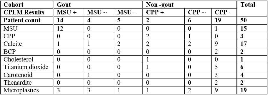
Results: A summary of the results from this experiment can be found in figure 3. We demonstrate a high correspondence (95%) between a certain identification with PLM and iRPolM. While the rheumatologist is only able to identify MSU/CPPD, Raman can also discriminate other microparticles.

Hydroxyapatite crystals were found in 1 patient with RA and one patient with a degenerative shoulder syndrome. Calcite (calcium carbonate) could be identified in 17 patients, 4 of whom were diagnosed with gout, 4 with CPPD, 3 with Rheumatoid Arthritis, 2 with Osteoarthritis and 4 undefined.

Titanium dioxide crystals were found in 6 patients. Only one of 6 patients with titanium crystals had an orthopedic implant, there is no clear relation between the two.

Carotenoids, a precursor molecule for vitamin A, could be found in 4 patients, 1 of which was diagnosed with gout and 3 had an undefined joint disease. All thenardite (sodium sulphate) crystals could be found in patients with osteoarthritis. Microplastics could be found in 19 patients, but environmental pollution must be excluded.

Conclusion: Raman spectroscopy has a significant added value to PLM, as it can not only can discriminate between MSU and CPPD but also identify a whole range of other crystals which might be related to potentially novel crystallopathies. In the future crystal identification in rheumatology may well have to rely on Raman based diagnostics.

Supporting image 1

Patient characteristics table. * CPDD: calcium pyrophosphate deposition disease ** Undefined means joint disease other than gout, CPPD, rheumatoid arthritis or osteoarthritis. *** Yes/no means a 100% certain crystal identification as assessed by an experienced rheumatologist. Doubtful means a less than 100% certain crystal identification

Supporting image 2

Graphic depiction of the Raman analysis process. After scanning, Raman spectra are analyzed with visual examination by a trained spectroscopist. The presence of a peak at 630 wavenumber (cm_1) is specific for MSU crystals, whereas a peak at 1050 wavenumber (cm_1) is specific for calcium pyrophosphate crystals.

Supporting image 3

Results of sample analysis. + represents certain positive identification, ~ represents uncertain, – represents certain negative identification. Numbers represent counts of patients with Raman detected crystals in this category (i.e. gout, MSU+ CPLM had 1 patient with calcite crystals).


Disclosures: T. Niessink, None; M. Janssen, None; C. Otto, Hybriscan BV; T. Jansen, None.

To cite this abstract in AMA style:

Niessink T, Janssen M, Otto C, Jansen T. Raman Spectroscopy Integrated with Polarized Light Microscopy for Diagnosis of Crystallopathies [abstract]. Arthritis Rheumatol. 2022; 74 (suppl 9). https://acrabstracts.org/abstract/raman-spectroscopy-integrated-with-polarized-light-microscopy-for-diagnosis-of-crystallopathies/. Accessed .
  • Tweet
  • Click to email a link to a friend (Opens in new window) Email
  • Click to print (Opens in new window) Print

« Back to ACR Convergence 2022

ACR Meeting Abstracts - https://acrabstracts.org/abstract/raman-spectroscopy-integrated-with-polarized-light-microscopy-for-diagnosis-of-crystallopathies/

Advanced Search

Your Favorites

You can save and print a list of your favorite abstracts during your browser session by clicking the “Favorite” button at the bottom of any abstract. View your favorites »

All abstracts accepted to ACR Convergence are under media embargo once the ACR has notified presenters of their abstract’s acceptance. They may be presented at other meetings or published as manuscripts after this time but should not be discussed in non-scholarly venues or outlets. The following embargo policies are strictly enforced by the ACR.

Accepted abstracts are made available to the public online in advance of the meeting and are published in a special online supplement of our scientific journal, Arthritis & Rheumatology. Information contained in those abstracts may not be released until the abstracts appear online. In an exception to the media embargo, academic institutions, private organizations, and companies with products whose value may be influenced by information contained in an abstract may issue a press release to coincide with the availability of an ACR abstract on the ACR website. However, the ACR continues to require that information that goes beyond that contained in the abstract (e.g., discussion of the abstract done as part of editorial news coverage) is under media embargo until 10:00 AM ET on November 14, 2024. Journalists with access to embargoed information cannot release articles or editorial news coverage before this time. Editorial news coverage is considered original articles/videos developed by employed journalists to report facts, commentary, and subject matter expert quotes in a narrative form using a variety of sources (e.g., research, announcements, press releases, events, etc.).

Violation of this policy may result in the abstract being withdrawn from the meeting and other measures deemed appropriate. Authors are responsible for notifying colleagues, institutions, communications firms, and all other stakeholders related to the development or promotion of the abstract about this policy. If you have questions about the ACR abstract embargo policy, please contact ACR abstracts staff at [email protected].

Wiley

  • Online Journal
  • Privacy Policy
  • Permissions Policies
  • Cookie Preferences

© Copyright 2025 American College of Rheumatology