ACR Meeting Abstracts

ACR Meeting Abstracts

  • Meetings
    • ACR Convergence 2024
    • ACR Convergence 2023
    • 2023 ACR/ARP PRSYM
    • ACR Convergence 2022
    • ACR Convergence 2021
    • ACR Convergence 2020
    • 2020 ACR/ARP PRSYM
    • 2019 ACR/ARP Annual Meeting
    • 2018-2009 Meetings
    • Download Abstracts
  • Keyword Index
  • Advanced Search
  • Your Favorites
    • Favorites
    • Login
    • View and print all favorites
    • Clear all your favorites
  • ACR Meetings

Abstract Number: 1314

Quantitative MRI Measurement of Tenosynovitis Demonstrates Differing Responses of Synovitis and Tenosynovitis after RA Treatment

Michael A. Bowes1, Gwenael Guillard1, Graham R. Vincent2, Jane E. Freeston3, Edward M. Vital4, Paul Emery5 and Philip G. Conaghan6, 1Imorphics, Manchester, United Kingdom, 2Imorphics Ltd, Manchester, United Kingdom, 3LIMM Section of Musculoskeletal Disease, University of Leeds, Leeds, United Kingdom, 4Leeds Institute of Rheumatic and Musculoskeletal Medicine, University of Leeds, Leeds, United Kingdom, 5Leeds Institute of Rheumatic and Musculoskeletal Medicine, Leeds Gen Infirmary, Leeds, United Kingdom, 6University of Leeds & NIHR Leeds Musculoskeletal Biomedical Research Unit, Leeds, United Kingdom

Meeting: 2015 ACR/ARHP Annual Meeting

Date of first publication: September 29, 2015

Keywords: Imaging, MRI, Rheumatoid arthritis (RA), synovitis and tendonitis/bursitis

  • Tweet
  • Email
  • Print
Session Information

Date: Monday, November 9, 2015

Title: Imaging of Rheumatic Diseases Poster II: X-ray, MRI, PET and CT

Session Type: ACR Poster Session B

Session Time: 9:00AM-11:00AM

Background/Purpose: Inflammation of the
tendon sheaths (tenosynovitis) is a recognised component of rheumatoid
arthritis (RA). A comprehensive assessment of inflammation will require the
inclusion of tenosynovitis as well as synovitis and osteitis, and consequently
it is proposed to include a semi-quantitative assessment of wrist tenosynovitis
in the OMERACT RAMRIS scoring system. 
Active appearance models (AAM) have been successfully used to develop an
automatic quantitative version of the current RAMRIS methodology. 

This
study was a pilot investigation in established RA to assess whether AAMs can be
used to produce an automatic tenosynovitis measure and compare the response to
therapy of quantitative wrist tenosynovitis and synovitis measures.

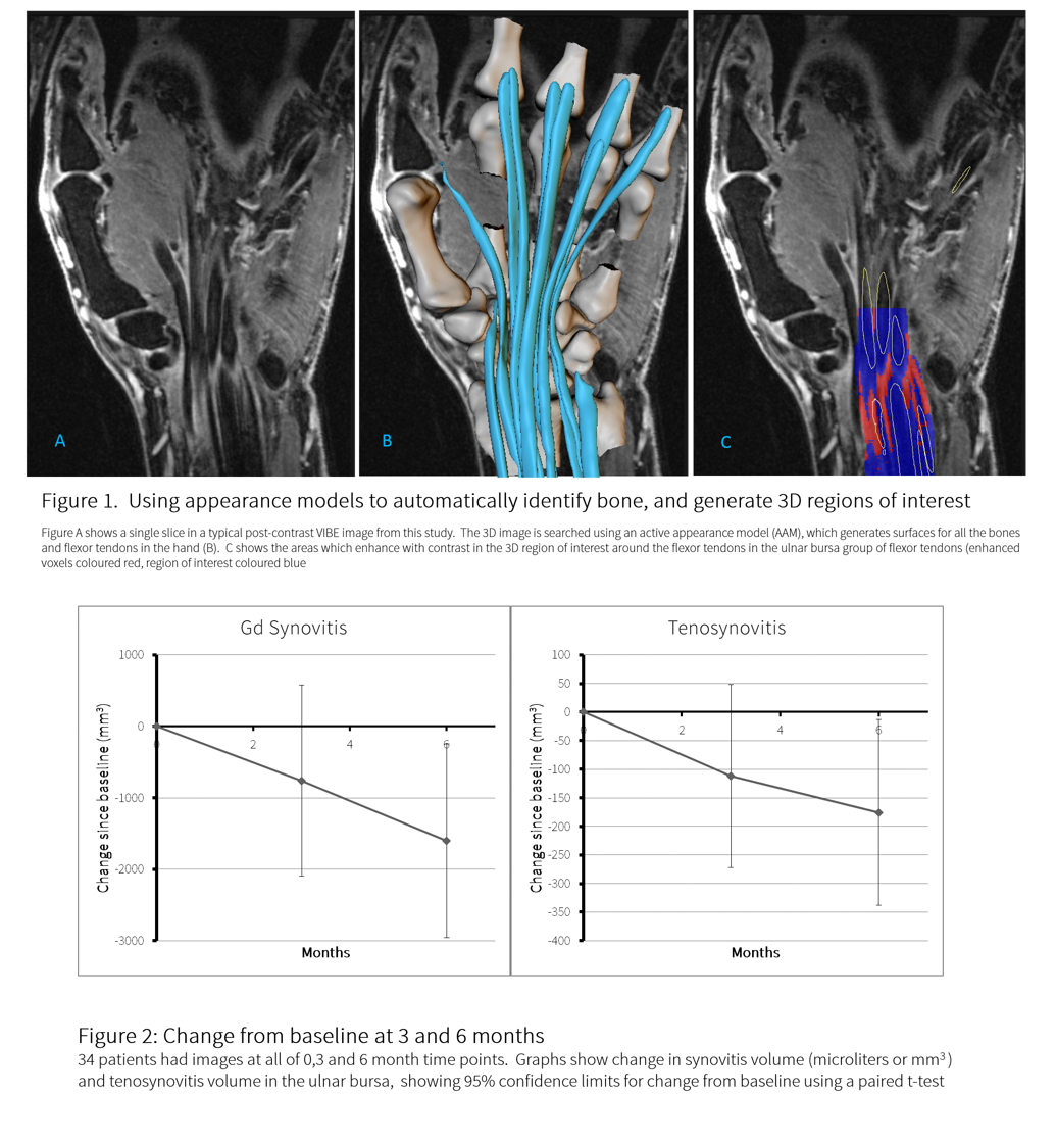
Methods: MR images of the hand were
acquired at 0, 3 and 6 months from 34 established, seropositive RA patients who
received a cycle of rituximab therapy in an open label study.  Pre- and post-contrast VIBE images with fat
saturation were acquired, and searched with AAMs to identify bones and capsular
structures and generate 3D regions of interest (ROIs).  Volume which enhanced with contrast was
calculated using a shuffle transform. AAMs of the flexor tendons were generated
from an independent training set of hand MR images.  Briefly, the process includes manual
segmentation of the tendons by an expert, generating 3D surfaces using a
marching cubes algorithm and the generation of AAMs for each tendon. Images
were automatically searched using the AAM, and visually inspected to ensure
that the search process had correctly identified the tendons.  A 3D region of interest (ROI) around each
tendon was created by inflating the tendon shape to form a halo which included
the tendon sheath.  Within the ROI the
tenosynovitis volume was calculated using the shuffle transform method. For
this pilot study only the wrist flexor tendons within the common synovial
sheath were analysed.  The amount of
change for the 2 methods was judged using a paired t-test.

Results:

Tenosynovitis
in the flexor tendons, and synovitis volume decreased at 3 and 6 months, in an
approximately linear fashion.  Change was
significant at 6 months for both measures (Figure 2). Although the change in
the population mean was linear for both measures,  the slope of change in tenosynovitis volume
for individual patients did not correlate with change in synovitis volume for
the same patients (r2 = 0.29).

Conclusion:

It
is feasible to quantify tenosynovitis using AAMs.  Tenosynovitis in the flexor tendons decreased
over 6 months, and only correlated weakly with change in synovitis volume
within the same patient. Tenosynovitis appears to be as responsive as synovial volume,
but did not correlate with synovial change in individual patients in this small
study.  Tenosynovitis may therefore add
new information to that already provided by measures of synovitis, though this
will need confirmation with a fully developed tool in a larger RA population.

ALT


Disclosure: M. A. Bowes, None; G. Guillard, None; G. R. Vincent, None; J. E. Freeston, Chugai, Pfizer, 5; E. M. Vital, Roche Pharmaceuticals, 2,Roche Pharmaceuticals, GSK, UCB, Chugai, 5; P. Emery, Abbvie, BMS, Lilly, MSD, Novartis, Pfizer, Roche, Samsung, Sandoz, UCB, 2,Abbvie, BMS, Lilly, MSD, Novartis, Pfizer, Roche, Samsung, Sandoz, UCB, 5; P. G. Conaghan, Abbvie, BMS, Novartis Non-remunerative, 5,Abbvie, BMS, Janssen, Roche, 8.

To cite this abstract in AMA style:

Bowes MA, Guillard G, Vincent GR, Freeston JE, Vital EM, Emery P, Conaghan PG. Quantitative MRI Measurement of Tenosynovitis Demonstrates Differing Responses of Synovitis and Tenosynovitis after RA Treatment [abstract]. Arthritis Rheumatol. 2015; 67 (suppl 10). https://acrabstracts.org/abstract/quantitative-mri-measurement-of-tenosynovitis-demonstrates-differing-responses-of-synovitis-and-tenosynovitis-after-ra-treatment/. Accessed .
  • Tweet
  • Email
  • Print

« Back to 2015 ACR/ARHP Annual Meeting

ACR Meeting Abstracts - https://acrabstracts.org/abstract/quantitative-mri-measurement-of-tenosynovitis-demonstrates-differing-responses-of-synovitis-and-tenosynovitis-after-ra-treatment/

Advanced Search

Your Favorites

You can save and print a list of your favorite abstracts during your browser session by clicking the “Favorite” button at the bottom of any abstract. View your favorites »

All abstracts accepted to ACR Convergence are under media embargo once the ACR has notified presenters of their abstract’s acceptance. They may be presented at other meetings or published as manuscripts after this time but should not be discussed in non-scholarly venues or outlets. The following embargo policies are strictly enforced by the ACR.

Accepted abstracts are made available to the public online in advance of the meeting and are published in a special online supplement of our scientific journal, Arthritis & Rheumatology. Information contained in those abstracts may not be released until the abstracts appear online. In an exception to the media embargo, academic institutions, private organizations, and companies with products whose value may be influenced by information contained in an abstract may issue a press release to coincide with the availability of an ACR abstract on the ACR website. However, the ACR continues to require that information that goes beyond that contained in the abstract (e.g., discussion of the abstract done as part of editorial news coverage) is under media embargo until 10:00 AM ET on November 14, 2024. Journalists with access to embargoed information cannot release articles or editorial news coverage before this time. Editorial news coverage is considered original articles/videos developed by employed journalists to report facts, commentary, and subject matter expert quotes in a narrative form using a variety of sources (e.g., research, announcements, press releases, events, etc.).

Violation of this policy may result in the abstract being withdrawn from the meeting and other measures deemed appropriate. Authors are responsible for notifying colleagues, institutions, communications firms, and all other stakeholders related to the development or promotion of the abstract about this policy. If you have questions about the ACR abstract embargo policy, please contact ACR abstracts staff at [email protected].

Wiley

  • Online Journal
  • Privacy Policy
  • Permissions Policies
  • Cookie Preferences

© Copyright 2025 American College of Rheumatology