Session Information
Date: Sunday, November 8, 2020
Title: Systemic Sclerosis & Related Disorders – Basic Science (1522–1526)
Session Type: Abstract Session
Session Time: 3:00PM-3:50PM
Background/Purpose: The radiological hallmarks of systemic sclerosis-related interstitial lung disease (SSc-ILD) include interstitial inflammation (ground glass opacity) with reticular changes (fibrosis). The precise pathobiology of these radiographic manifestations is unknown. We hypothesized that certain cytokines, chemokines and growth factors may uniquely associate with distinct radiographic patterns of ILD in SSc.
Methods: We measured the concentrations of 68 chemokines, cytokines and growth factors from bronchoalveolar lavage (BAL) fluid of 103 patients with SSc-ILD who participated in Scleroderma Lung Study I (1). SLS I randomized SSc-ILD participants to 1 year of oral cyclophosphamide versus placebo, followed by 1 year off all treatment. Bronchoscopy was performed at baseline in SLS I. Quantitative imaging analysis (QIA) was used to calculate the extent of radiographic fibrosis (QLF) and ground glass opacity (GGO) on the baseline HRCT of the chest. Kendall tau’s correlations were calculated to examine the relationship between BAL proteins and QLF and GGO scores, separately, for the whole lung. Multivariable linear regression models were created to determine the key BAL proteins associated with QLF and GGO scores using a backward selection process in combination with evaluation of the Akaike information criterion (AIC). The bootstrap procedure was employed for internal validation.
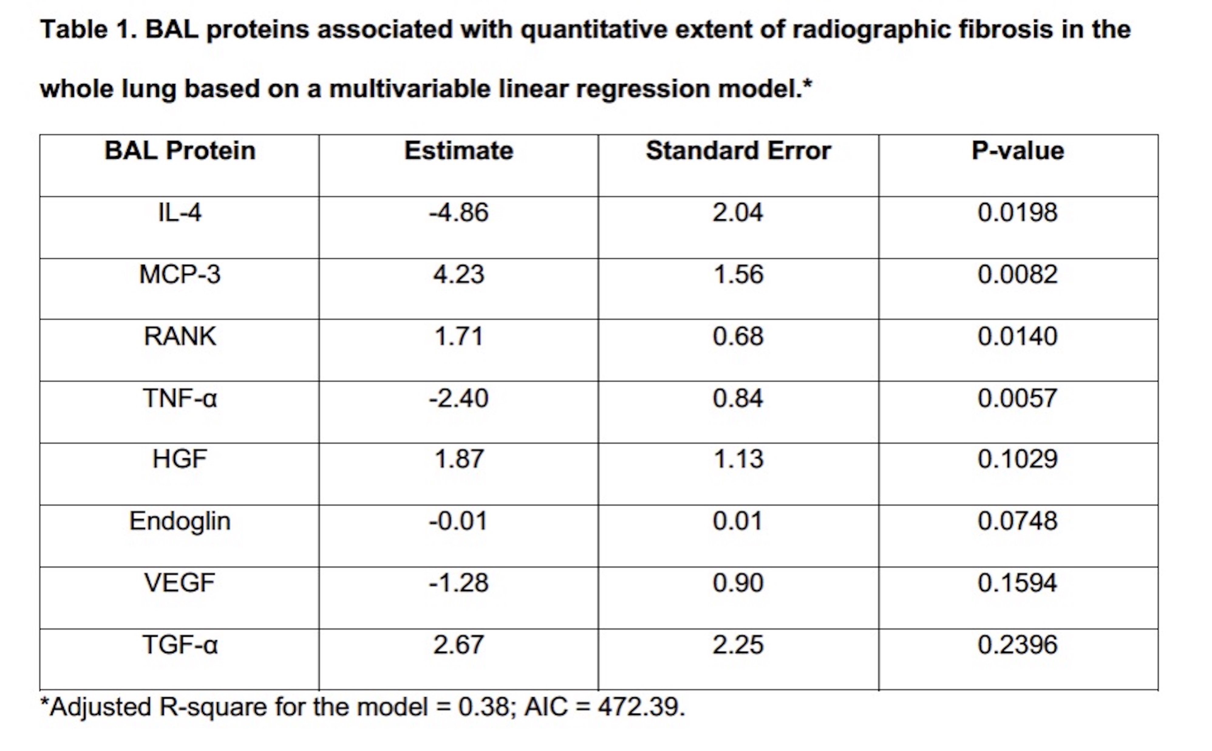
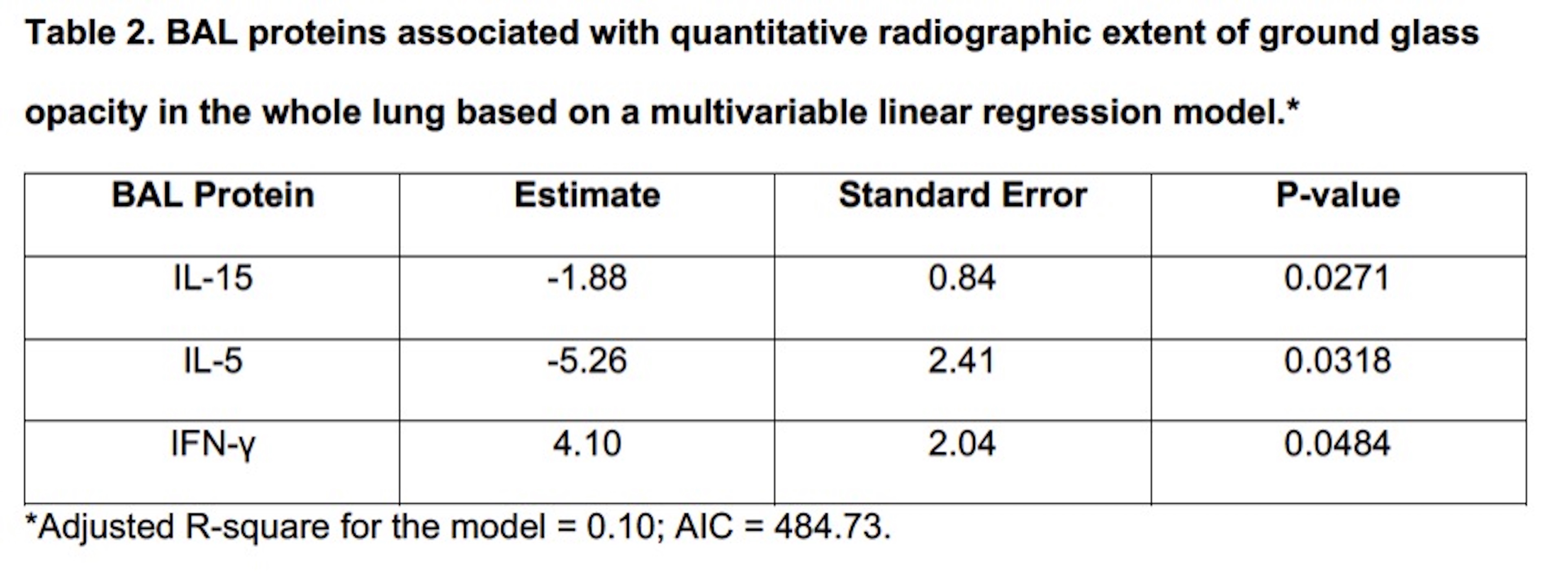
Results: QLF scores correlated significantly with 25 different proteins from several biologic pathways including pro-fibrotic factors (transforming growth factor beta [TGF-β], platelet-derived growth factor [PDGF]), proteins involved in tissue remodeling (Matrix metallopeptidase [MMP]-1,7,8,9; Hepatocyte growth factor [HGF]), and those involved in monocyte/macrophage migration and activation (Monocyte chemoattractant protein [MCP]-1,3; macrophage colony-stimulating factor [MCSF]). GGO scores correlated with 7 different proteins that are mediators of immune response and inflammation (interleukin [IL]-5, IL-15, IL-1 receptor antagonist and interferon gamma [IFN-γ]) with limited overlap to proteins that related to fibrosis. Vascular endothelial growth factor (VEGF) levels were lower in patients with more extensive GGO and QLF, suggesting that vascular changes are an important feature of SSc-ILD. In the multivariate models, IL-4, MCP-3, receptor activator of NF-κB (RANK), tumor necrosis factor alpha (TNF-α) were independently associated with QLF (Table 1); whereas, IL-15, IL-5 and IFN-γ were independently associated with GGO (Table 2).
Conclusion: In a diverse and comprehensive analysis of BAL proteins from a well-characterized SSc-ILD cohort, the findings suggest that specific biological signatures underlie distinct radiographic features of ILD in SSc. These proteins may represent important therapeutic targets and serve to help define specific phenotypes of SSc-ILD (inflammation predominant versus fibrosis predominant), which may ultimately lead to the development of personalized treatment approaches in this patient population.
To cite this abstract in AMA style:
Volkmann E, Tashkin D, Li N, Leng M, Kim G, Goldin J, Harui A, Roth M. Pulmonary Cytokine, Chemokine and Growth Factor Profiles of Distinct Radiographic Patterns of Interstitial Lung Disease in Systemic Sclerosis [abstract]. Arthritis Rheumatol. 2020; 72 (suppl 10). https://acrabstracts.org/abstract/pulmonary-cytokine-chemokine-and-growth-factor-profiles-of-distinct-radiographic-patterns-of-interstitial-lung-disease-in-systemic-sclerosis/. Accessed .« Back to ACR Convergence 2020
ACR Meeting Abstracts - https://acrabstracts.org/abstract/pulmonary-cytokine-chemokine-and-growth-factor-profiles-of-distinct-radiographic-patterns-of-interstitial-lung-disease-in-systemic-sclerosis/


