Session Information
Session Type: ACR Poster Session C
Session Time: 9:00AM-11:00AM
Background/Purpose:
Systemic Lupus Erythematosus (SLE) patients display high levels of
the cell-bound complement activation factor C4d deposits on erythrocytes, B
lymphocytes and platelets. In particular, complement activation on platelets
has been suggested to play a crucial role in the pathogenesis of thrombosis in
SLE, especially in aPL positive patients. Several in vitro and in vivo studies
have demonstrated an increased C4d deposition on platelets, that have been
associated with stroke or other thrombotic complications in SLE.
Recently, the addition of purified anti-cardiolipin antibodies
from Antiphospholipid antibody syndrome (APS) patients has been demonstrated to
increase C4d deposition on activated fixed platelets.
Thrombosis is an hallmark of APS and both platelets and complement
have been suggested to be involved in the pathogenic pathways. However few data
on complement activation and C4d deposition on platelets in Primary APS
patients have been reported in the literature. Aim of the study was to evaluate
C4d deposition on platelets, erythrocytes and B lymphocytes of Primary APS
patients (PAPS) in comparison to aPL neg SLE patients and healthy donors (NHS).
Methods:
11 active SLE (SLEDAI 3-14) and 18 PAPS patients attending our
Rheumatology Unit were consecutively enrolled. C4d deposition on platelets,
erythrocytes and B lymphocytes was assessed by flow cytometry, using an
anti-human C4d monoclonal antibody associated with specific lineage markers.
Results were expressed as C4d positive cell percentage, mean and Standard Error
Mean. Statistical analysis was performed by Mann Whitney test with significant
limit p<0.05.
Results:
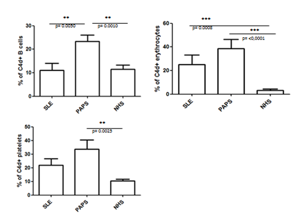
The proportion of C4d positive cells was higher in PAPS than SLE
in all cell populations, with statistical significance for B cells (p=0.005)
(fig 1). Comparing PAPS vs NHS, significant differences were detected for all
cell types (p<0.005), while only erythrocytes C4d deposits were
significantly higher in SLE patients than in NHS.
Conclusion:
we show for the first time that PAPS patients display higher
levels of cell bound C4d in comparison to both NHS and SLE. These in vivo data
suggest that: i) complement is activated in PAPS in spite of normal C3/C4
plasma levels: ii) increased complement activation on platelets may play a role
in causing thrombocytopenia in PAPS. The increased C4d deposition on B
lymphocytes deserves further investigation. As a whole our finding does suggest
that cell-bound complement split products may represent a potential tool to
characterize APS clinical subtypes.
Figure 1: Cd4 positive cells in PAPS
and SLE
To cite this abstract in AMA style:
Gerosa M, Lonati PA, Ubiali T, Cornalba M, Borghi MO, Meroni PL. Primary Antiphospholipid Syndrome Patients Display Increased Levels of Cell-Bound C4d in Comparison to SLE and Healthy Donors [abstract]. Arthritis Rheumatol. 2015; 67 (suppl 10). https://acrabstracts.org/abstract/primary-antiphospholipid-syndrome-patients-display-increased-levels-of-cell-bound-c4d-in-comparison-to-sle-and-healthy-donors/. Accessed .« Back to 2015 ACR/ARHP Annual Meeting
ACR Meeting Abstracts - https://acrabstracts.org/abstract/primary-antiphospholipid-syndrome-patients-display-increased-levels-of-cell-bound-c4d-in-comparison-to-sle-and-healthy-donors/

