Session Information
Session Type: Poster Session A
Session Time: 10:30AM-12:30PM
Background/Purpose: Juvenile idiopathic arthritis (JIA) is the most common chronic rheumatological disease in children less than 16 years of age. Various factors have been suggested to negatively influence JIA patients’ bone mineral density (BMD). Decreased BMD has a long-term implication in such patients, making them more prone to fractures later in life.
To determine the prevalence and risk factors associated with low bone mineral density (BMD) in children with Juvenile Idiopathic Arthritis (JIA).
Methods: This cross-sectional study was conducted at the Pediatric Rheumatology Clinic, Department of Pediatrics, All India Institute of Medical Sciences (AIIMS), New Delhi.
All children diagnosed with JIA as per ILAR criteria were screened for eligibility. Children aged 5-18 years with JIA and disease more than one year were enrolled; those with bisphosphonate intake, bone damage due to other causes like infection, trauma, and JIA with overlap syndrome were excluded. Demographic details, clinical assessments, and laboratory investigations, including serum calcium, CRP, RF/anti-CCP, vitamin D3, and intact PTH, were recorded in a predesigned performa. Disease activity and functional disability were assessed using the clinical Juvenile Arthritis Disease Activity Score-10 (cJADAS-10) and Steinbrocker. BMD was measured using DXA at the lumbar spine and left femoral neck. Low BMD was defined as a Z-score ≤ – 2 SD at either of the two sites. Analysis was done using StataCorp. 2021. Stata statistical software: Release 18. Multivariate logistic regression was done to identify factors associated with low BMD.
Results: 101 children with 68.3% (69/101) males with a mean age of 154.6 months with a median disease duration of 48 months and a median delay in initiation of therapy of 14 months. Enthesitis-related arthritis (54.5%) was the most common subtype, followed by systemic JIA (36%). The demographic and laboratory characteristics are depicted in Table 1.
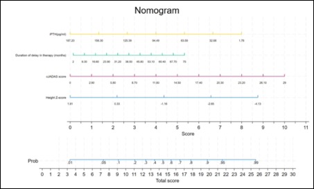
The prevalence of low BMD was 55.4% (56/101). Significant risk factors for low BMD included higher cJADAS scores, lower height Z-scores, longer delay in initiation of therapy, and higher levels of intact PTH. Univariate analysis identified BMI, disease duration, systemic steroid use, and CRP levels as potential risk factors. Multivariate analysis confirmed that lower height Z-scores and higher cJADAS scores were independently associated with low BMD. Table 2 depicts the results of the regression analysis. The area under the ROC curve was 0.79. A nomogram to predict the probability of low BMD was developed and is represented in Figure 1.
Conclusion: Significant children with JIA have low BMD. Addressing the identified risk factors, i.e. disease activity and delay in initiation of therapy, might help prevent long-term skeletal complications.
To cite this abstract in AMA style:
bagri n, Kalsotra S, jain v, Jana M, Khan M, Gupta Y, Das S. Prevalence and Risk Factors of Low Bone Mineral Density in Children with Juvenile Idiopathic Arthritis [abstract]. Arthritis Rheumatol. 2024; 76 (suppl 9). https://acrabstracts.org/abstract/prevalence-and-risk-factors-of-low-bone-mineral-density-in-children-with-juvenile-idiopathic-arthritis/. Accessed .« Back to ACR Convergence 2024
ACR Meeting Abstracts - https://acrabstracts.org/abstract/prevalence-and-risk-factors-of-low-bone-mineral-density-in-children-with-juvenile-idiopathic-arthritis/


