ACR Meeting Abstracts

ACR Meeting Abstracts

  • Meetings
    • ACR Convergence 2024
    • ACR Convergence 2023
    • 2023 ACR/ARP PRSYM
    • ACR Convergence 2022
    • ACR Convergence 2021
    • ACR Convergence 2020
    • 2020 ACR/ARP PRSYM
    • 2019 ACR/ARP Annual Meeting
    • 2018-2009 Meetings
    • Download Abstracts
  • Keyword Index
  • Advanced Search
  • Your Favorites
    • Favorites
    • Login
    • View and print all favorites
    • Clear all your favorites
  • ACR Meetings

Abstract Number: 1044

Prevalence and Distribution of Sonographic Elementary Lesions in PsA – Results of 2 Cohorts

Ari Polachek1, Victoria Furer2, Sydney Thib3, Liran Mendel2, Ori Elkayam2 and Lihi Eder4, 1Sourasky Medical Center, Tel Aviv, Israel, 2Tel Aviv Medical Center, Tel Aviv, Israel, 3Women’s College Research Institute, Toronto, ON, Canada, 4Women’s College Research Institute, Division of Rheumatology, University of Toronto, Toronto, ON, Canada

Meeting: ACR Convergence 2023

Keywords: Disease Activity, Imaging, Inflammation, Psoriatic arthritis, Ultrasound

  • Tweet
  • Click to email a link to a friend (Opens in new window) Email
  • Click to print (Opens in new window) Print
Session Information

Date: Monday, November 13, 2023

Title: (1033–1051) Imaging of Rheumatic Diseases Poster I

Session Type: Poster Session B

Session Time: 9:00AM-11:00AM

Background/Purpose: Psoriatic arthritis (PsA) can manifest with different musculoskeletal (MSK) features. Ultrasound (US) optimizes the assessment of the different MSK features in PsA. However, there is no consensus on which MSK sonographic lesions and what locations should be evaluated by US. The group for research and assessment of psoriasis and psoriatic arthritis (GRAPPA) have set a goal of defining the most prevalent locations for the various US lesions in order to reduce the number of scanned sites.

The aim of the study was to describe the prevalence and distribution of key sonographic MSK lesions in patients with PsA.

Methods: This study analyzed data from two prospectively recruited PsA cohorts. Cohort 1 included 101 patients with active PsA (DAPSA >14) and cohort 2 included 94 patients with active PsA prior to initiation of therapy. All underwent a comprehensive US assessment, including both gray scale (GS) and power Doppler (PD) of 50 joints, 40 tendons and 14 entheses. The following sonographic lesions were assessed by two sonographers blinded to clinical data: I. Inflammatory lesions – synovitis, tenosynovitis, peritenonitis and enthesitis and II. Structural lesions – periarticular erosions and bone proliferations. Presence or Absence of these lesions was determined based on previously suggested definitions by OMERACT (when available) or other publication and their prevalence by joint/tendon site was reported.

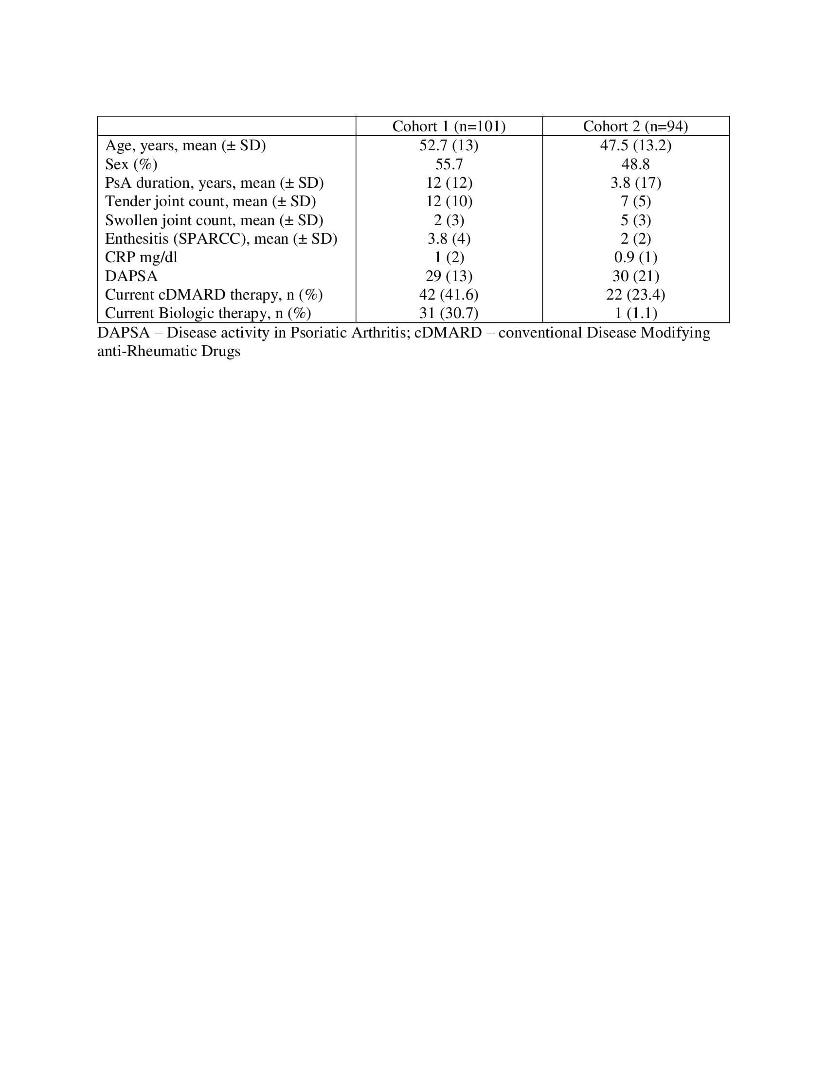
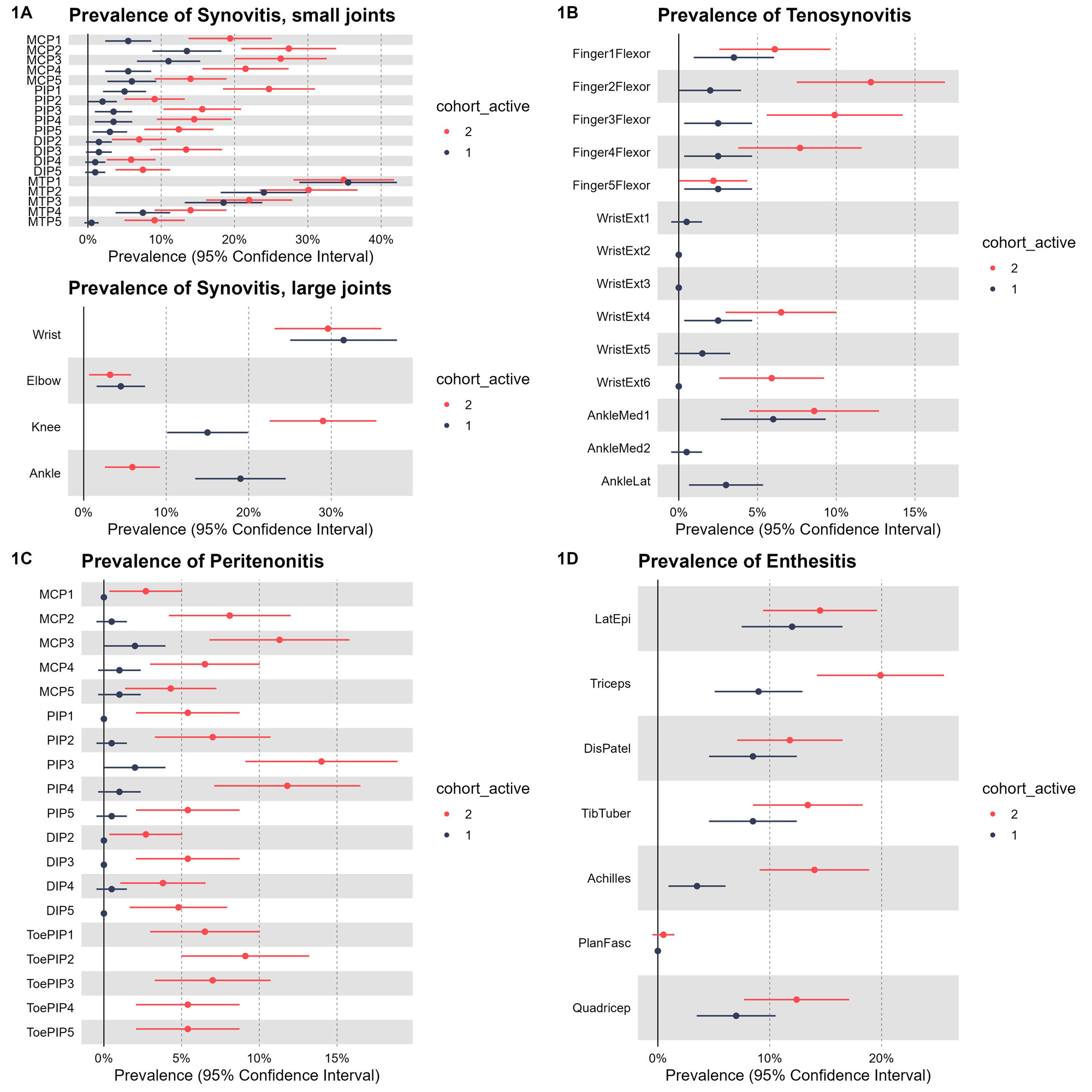
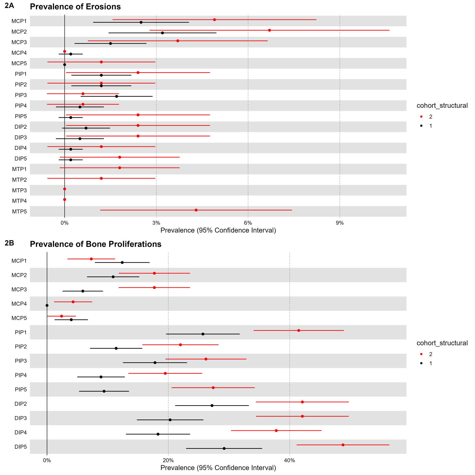
Results: In cohort 1, mean ± SD age was 52.7 ± 13 and 55.7% were females. In cohort 2, mean ± SD age was 47.5 ± 13.2 and 48.8% were females (Table 1). The most prevalent locations of the inflammatory lesions in both cohorts were (figure 1): Synovitis (small joints) – MCP2 (cohort 1: 13.5%, cohort 2: 27%), MCP 3 (cohort 1: 11%, cohort 2: 26%), IP1 (cohort 1: 5%, cohort 2: 24.5%) PIP 3 (cohort 1:3%, cohort 2: 16%), MTP 1 (cohort 1: 35.5%, cohort 2: 35%), MTP2 (cohort 1: 24%, cohort 2: 30%), MTP3 (cohort 1: 18.5%, cohort 2: 22%); Synovitis (medium-large joints) – wrist (cohort 1: 31.5%, cohort 2: 29%) and knee (cohort 1: 15%, cohort 2: 29%); Tenosynovitis – 2nd finger flexors (cohort 1: 2%, cohort 2: 8%) 3rd finger flexor (cohort 1: 2%, cohort 2: 6%), extensor digitorum (cohort 1: 2.5%, cohort 2: 6%), tibialis posterior (cohort 1: 6%, cohort 2: 8.5%); extensor peritenonitis –MCP3 (cohort 1: 2%, cohort 2: 12%), PIP 3 (cohort 1: 2%, cohort 2: 14%), PIP4 (cohort 1: 1%, cohort 2: 12%); Enthesitis – lateral epicondyle (cohort 1: 12%, cohort 2: 14%) and triceps (cohort 1: 9%, cohort 2: 20%). The most prevalent locations of the structural lesions (figure 2): Erosions – MCP1 (cohort 1: 4%, cohort 2: 4%), MCP2 (cohort 1: 6.5%, cohort 2: 6%), MCP3 (cohort 1: 3%, cohort 2: 3%), MTP5 (cohort 2: 4%); Bone proliferations – IP1 (cohort 1: 25%, cohort 2: 36%), DIP2 (cohort 1: 27.5%, cohort 2: 37%), DIP3 (cohort 1: 19.5%, cohort 2: 37%) and DIP5 (cohort 1: 28%, cohort 2: 44%).

Conclusion: This descriptive study provides comprehensive information on the most commonly affected sites for key inflammatory and structural domains in PsA. This information can inform efforts to develop reduced sonographic score to diagnose or monitor disease activity in PsA.

Supporting image 1

Table 1: Baseline and clinical characteristics

Supporting image 2

Figure1: Prevalence and distribution of sonographic inflammatory lesions in PsA

Supporting image 3

Figure2: Prevalence and distribution of sonographic structural lesions in PsA


Disclosures: A. Polachek: None; V. Furer: None; S. Thib: None; L. Mendel: None; O. Elkayam: None; L. Eder: AbbVie, 2, 5, Eli Lilly, 2, 5, Fresenius Kabi, 5, Janssen, 2, 5, Novartis, 2, 5, Pfizer Inc, 2, 5, Sandoz, 5, UCB, 2, 5.

To cite this abstract in AMA style:

Polachek A, Furer V, Thib S, Mendel L, Elkayam O, Eder L. Prevalence and Distribution of Sonographic Elementary Lesions in PsA – Results of 2 Cohorts [abstract]. Arthritis Rheumatol. 2023; 75 (suppl 9). https://acrabstracts.org/abstract/prevalence-and-distribution-of-sonographic-elementary-lesions-in-psa-results-of-2-cohorts/. Accessed .
  • Tweet
  • Click to email a link to a friend (Opens in new window) Email
  • Click to print (Opens in new window) Print

« Back to ACR Convergence 2023

ACR Meeting Abstracts - https://acrabstracts.org/abstract/prevalence-and-distribution-of-sonographic-elementary-lesions-in-psa-results-of-2-cohorts/

Advanced Search

Your Favorites

You can save and print a list of your favorite abstracts during your browser session by clicking the “Favorite” button at the bottom of any abstract. View your favorites »

All abstracts accepted to ACR Convergence are under media embargo once the ACR has notified presenters of their abstract’s acceptance. They may be presented at other meetings or published as manuscripts after this time but should not be discussed in non-scholarly venues or outlets. The following embargo policies are strictly enforced by the ACR.

Accepted abstracts are made available to the public online in advance of the meeting and are published in a special online supplement of our scientific journal, Arthritis & Rheumatology. Information contained in those abstracts may not be released until the abstracts appear online. In an exception to the media embargo, academic institutions, private organizations, and companies with products whose value may be influenced by information contained in an abstract may issue a press release to coincide with the availability of an ACR abstract on the ACR website. However, the ACR continues to require that information that goes beyond that contained in the abstract (e.g., discussion of the abstract done as part of editorial news coverage) is under media embargo until 10:00 AM ET on November 14, 2024. Journalists with access to embargoed information cannot release articles or editorial news coverage before this time. Editorial news coverage is considered original articles/videos developed by employed journalists to report facts, commentary, and subject matter expert quotes in a narrative form using a variety of sources (e.g., research, announcements, press releases, events, etc.).

Violation of this policy may result in the abstract being withdrawn from the meeting and other measures deemed appropriate. Authors are responsible for notifying colleagues, institutions, communications firms, and all other stakeholders related to the development or promotion of the abstract about this policy. If you have questions about the ACR abstract embargo policy, please contact ACR abstracts staff at [email protected].

Wiley

  • Online Journal
  • Privacy Policy
  • Permissions Policies
  • Cookie Preferences

© Copyright 2025 American College of Rheumatology