ACR Meeting Abstracts

ACR Meeting Abstracts

  • Meetings
    • ACR Convergence 2024
    • ACR Convergence 2023
    • 2023 ACR/ARP PRSYM
    • ACR Convergence 2022
    • ACR Convergence 2021
    • ACR Convergence 2020
    • 2020 ACR/ARP PRSYM
    • 2019 ACR/ARP Annual Meeting
    • 2018-2009 Meetings
    • Download Abstracts
  • Keyword Index
  • Advanced Search
  • Your Favorites
    • Favorites
    • Login
    • View and print all favorites
    • Clear all your favorites
  • ACR Meetings

Abstract Number: 1285

PET/CT Imaging in Patients with Vascular Behcet Disease

Bahtiyar Toz1, Burak Erer1, Sevil Kamali1, Murat Inanc2, Lale Ocal3 and Ahmet Gul1, 1Department of Internal Medicine, Division of Rheumatology, Istanbul University, Istanbul Faculty of Medicine, Istanbul, Turkey, 2Department of Internal Medicine, Division of Rheumatology, Istanbul University, Istanbul Faculty of Medicine, İstanbul, Turkey, 3Department of Internal Medicine, Rheumatology Division, Istanbul University, Istanbul Faculty of Medicine, Istanbul, Turkey

Meeting: 2016 ACR/ARHP Annual Meeting

Date of first publication: September 28, 2016

Keywords: Behcet's syndrome

  • Tweet
  • Email
  • Print
Session Information

Date: Monday, November 14, 2016

Title: Imaging of Rheumatic Diseases - Poster II: XR/CT/PET/MRI

Session Type: ACR Poster Session B

Session Time: 9:00AM-11:00AM

Background/Purpose: BehetÕs disease (BD) is a multisystemic disease characterized by recurrent inflammatory manifestations. BD is classified as variable vessel vasculitis. While inflammatory thrombotic venous findings are dominant in patients with vascular-BD, arterial lesions manifesting as aneurysms and occlusive/stenotic lesions can also be seen.  In addition to clinical findings, imaging studies with CT, MRI, and Doppler ultrasonography play an important role in the diagnosis of vascular involvement. PET/CT is a relatively new tool being used in the assessment of large-vessel vasculitis, and there are conflicting reports about using PET/CT in patients with BD. We herein aimed to investigate our records for the role of PET-CT in the diagnosis and follow-up of BD patients with vascular involvement.

Methods:   We retrospectively reviewed the charts of BD patients who were investigated with PET/CT for any reason related to disease activity. Patients fulfilling the ISG criteria or with a preliminary diagnosis of BD were included for the analysis. Using a standard form, clinical findings, acute phase response including ESR and CRP, and additional imaging findings, such as CT or MRI, performed within the last 2-week of PET/CT scanning were recorded. Vascular FDG uptake was graded using a 4-point semi-quantitative scale. PET/CT scans were considered positive if vascular FDG uptake was ³ 2 (equal to or greater than liver).

Results: We identified 12 patients investigated with PET/CT. The mean age of the patients was 44 years, the mean disease duration was 14 years, and 11 (92%) were male. Demographic and clinical findings are summarized in Table 1. Patients underwent PET-CT due to fever of unknown origin (n=6), fatigue with unexplained high acute phase response (n=3), abdominal pain (n=1), or unexplained neck pain (n=1). Five of them fulfilled the ISG criteria, and 4 had positive PET/CT findings due to aortic involvement or bronchiolitis obliterans organizing pneumonia (n=2). No FDG uptake was detected in one patient with venous lesions. In remaining 7 patients with incomplete manifestations suggesting BD, vascular involvement documented by FDG uptake in aorta and its branches (n=2), pulmonary arteries (n=2), carotid arteries (n=2) and splenic artery (n=1). Description: Macintosh HD:Users:bahtiyartoz:Desktop:PAPER 2015:biten poster ve yazõlar:acr 2016:BH table.jpg

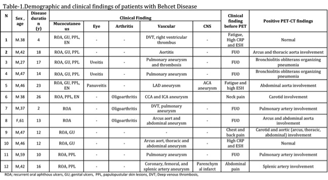
Conclusion: In BD patients with unexplained acute phase response, screening for vascular involvement is important, and PET/CT may contribute to diagnostic process by documenting medium-large size arterial activity. FDG uptake by arterial aneurysms and venous involvement in PET/CT is not clear, and several factors such as the size of the vessels and the thickness of vessel wall may affect FDG uptake. Parenchymal lesions possibly induced by small vessel vasculitis may also be another reason for positive PET/CT findings. Role of PET/CT in the diagnosis of patients with incomplete BD manifestations needs to be investigated further, since other disorders with mucocutaneous and vascular findings may mimic BD and cause diagnostic uncertainty.


Disclosure: B. Toz, None; B. Erer, None; S. Kamali, None; M. Inanc, None; L. Ocal, None; A. Gul, None.

To cite this abstract in AMA style:

Toz B, Erer B, Kamali S, Inanc M, Ocal L, Gul A. PET/CT Imaging in Patients with Vascular Behcet Disease [abstract]. Arthritis Rheumatol. 2016; 68 (suppl 10). https://acrabstracts.org/abstract/petct-imaging-in-patients-with-vascular-behcet-disease/. Accessed .
  • Tweet
  • Email
  • Print

« Back to 2016 ACR/ARHP Annual Meeting

ACR Meeting Abstracts - https://acrabstracts.org/abstract/petct-imaging-in-patients-with-vascular-behcet-disease/

Advanced Search

Your Favorites

You can save and print a list of your favorite abstracts during your browser session by clicking the “Favorite” button at the bottom of any abstract. View your favorites »

All abstracts accepted to ACR Convergence are under media embargo once the ACR has notified presenters of their abstract’s acceptance. They may be presented at other meetings or published as manuscripts after this time but should not be discussed in non-scholarly venues or outlets. The following embargo policies are strictly enforced by the ACR.

Accepted abstracts are made available to the public online in advance of the meeting and are published in a special online supplement of our scientific journal, Arthritis & Rheumatology. Information contained in those abstracts may not be released until the abstracts appear online. In an exception to the media embargo, academic institutions, private organizations, and companies with products whose value may be influenced by information contained in an abstract may issue a press release to coincide with the availability of an ACR abstract on the ACR website. However, the ACR continues to require that information that goes beyond that contained in the abstract (e.g., discussion of the abstract done as part of editorial news coverage) is under media embargo until 10:00 AM ET on November 14, 2024. Journalists with access to embargoed information cannot release articles or editorial news coverage before this time. Editorial news coverage is considered original articles/videos developed by employed journalists to report facts, commentary, and subject matter expert quotes in a narrative form using a variety of sources (e.g., research, announcements, press releases, events, etc.).

Violation of this policy may result in the abstract being withdrawn from the meeting and other measures deemed appropriate. Authors are responsible for notifying colleagues, institutions, communications firms, and all other stakeholders related to the development or promotion of the abstract about this policy. If you have questions about the ACR abstract embargo policy, please contact ACR abstracts staff at [email protected].

Wiley

  • Online Journal
  • Privacy Policy
  • Permissions Policies
  • Cookie Preferences

© Copyright 2025 American College of Rheumatology