ACR Meeting Abstracts

ACR Meeting Abstracts

  • Meetings
    • ACR Convergence 2024
    • ACR Convergence 2023
    • 2023 ACR/ARP PRSYM
    • ACR Convergence 2022
    • ACR Convergence 2021
    • ACR Convergence 2020
    • 2020 ACR/ARP PRSYM
    • 2019 ACR/ARP Annual Meeting
    • 2018-2009 Meetings
    • Download Abstracts
  • Keyword Index
  • Advanced Search
  • Your Favorites
    • Favorites
    • Login
    • View and print all favorites
    • Clear all your favorites
  • ACR Meetings

Abstract Number: 1553

Optical Tomography Can Accurately Diagnose Lupus Arthritis

Wei Tang1, Alessandro Marone2, Youngwan Kim2, Tommy Chen1, Cathy Guo1, Leila Khalili1, Andreas Hielscher2 and Anca Askanase3, 1Columbia University Medical Center, New York, NY, 2Columbia University, New York, NY, 3Columbia University College of Physicians & Surgeons, New York, NY

Meeting: ACR Convergence 2020

Keywords: Imaging, Measurement Instrument, pain, Systemic lupus erythematosus (SLE), Ultrasound

  • Tweet
  • Click to email a link to a friend (Opens in new window) Email
  • Click to print (Opens in new window) Print
Session Information

Date: Monday, November 9, 2020

Title: Imaging of Rheumatic Diseases Poster

Session Type: Poster Session D

Session Time: 9:00AM-11:00AM

Background/Purpose: Incapacitating inflammatory arthritis occurs in up to 88% of patients with systemic lupus erythematosus (SLE). Arthritis is present in 70-80% of lupus patients in clinical trials. There is continued debate as to whether tenderness in the absence of swelling constitutes active lupus arthritis and should be scored on activity instruments. The confusion over arthritis scoring impacts clinical care and trials. There is an unmet need for a simple tool that can objectively assess lupus arthritis; near-infrared optical imaging has the potential to address this need. Over the last decade the technology has been optimized for use in brain imaging, breast cancer, and peripheral ischemia. Studies have also shown its utility in rheumatoid arthritis. Near-infrared light illuminates the tissues and transmitted and reflected light intensities are measured. Maps computed from these measurements show the spatial distribution of physiological parameters. Changes in optical properties caused by physiologic changes form the basis for its diagnostic capabilities. Using a so-called frequency-domain optical imaging (FDOI) system1 we evaluated the proximal interphalangeal (PIP) joints of 16 SLE patients and 10 healthy volunteers.

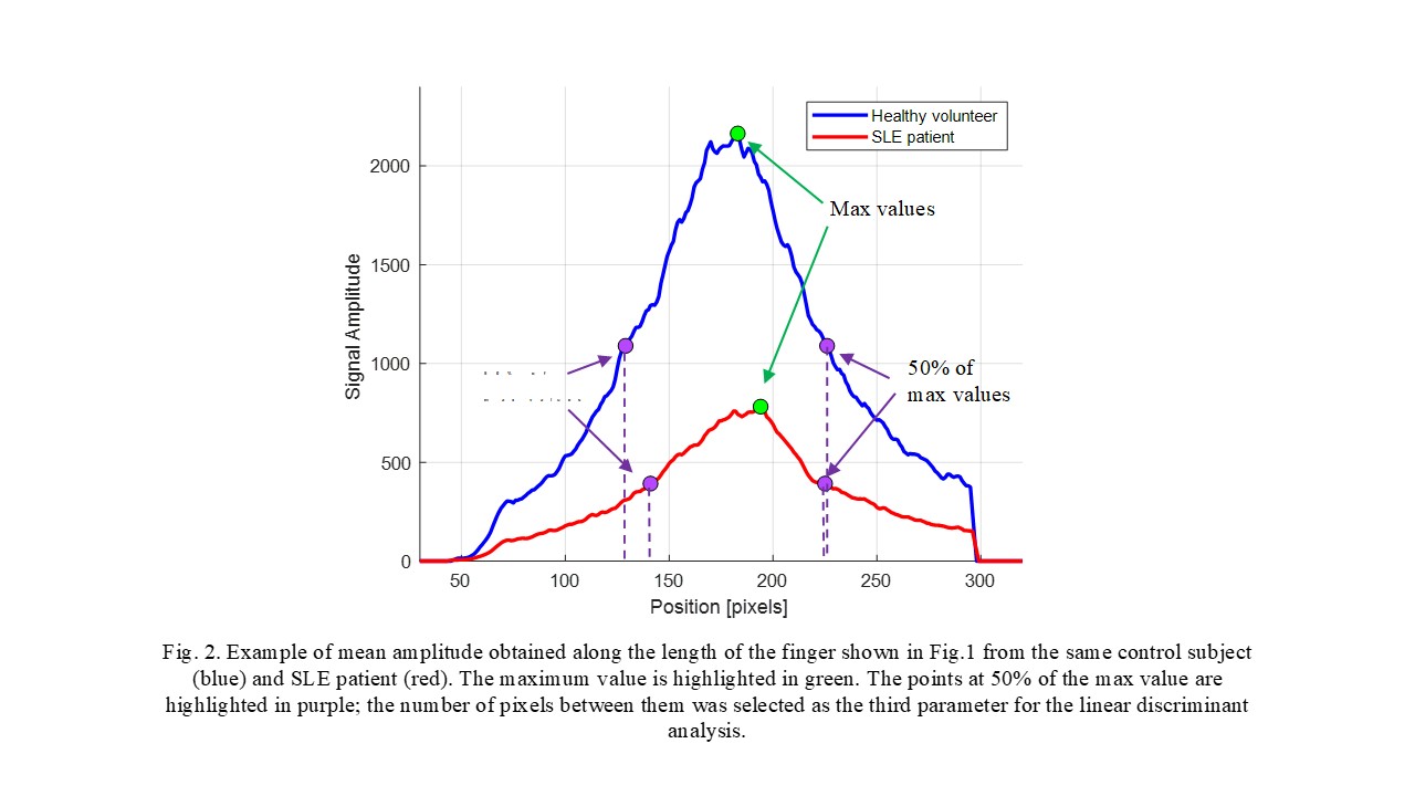
Methods: The FDOI system consists of a laser diode projecting a 1-mm – 670nm optical beam at 11 different positions along the PIP joints, 2mm apart, lengthwise. Fig.1 shows with the positions of the source. At each point, 16 images are acquired at different phases by a camera. The FDOI system is operated at a frequency of 300 MHz. PIPs 2-4 bilaterally were evaluated. A total of 60 healthy joints from 10 healthy volunteers and 96 joints from 16 patients with SLE arthritis were analyzed. Amplitudes and phases signals were generated using a demodulation algorithm.1 The fingers’ sizes (width and thickness) were measured using a caliper.

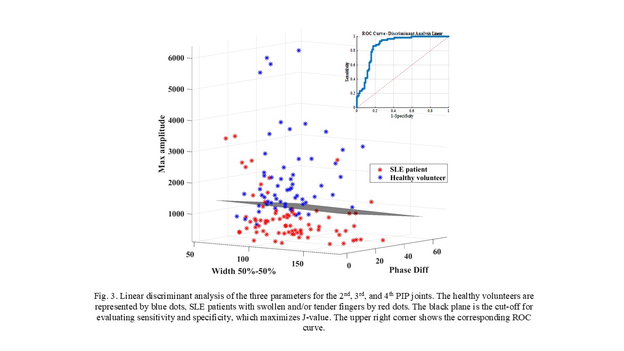
Results: The 16 patients with SLE arthritis (94% women, mean age 43±10 years, disease duration 14±8 years) had 74 swollen and/or tender PIP joints. Considering the mean signals from the 11 illumination positions (see Figs. 1 and 2), a discriminant analysis was done considering three variables: the maximum amplitude at the peak of the curve obtained, the maximum difference of the phase signals, and the number of pixels between 50% of the maximum value on the left and on the right of the peak. A receiver-operating curve (ROC) analysis showed an area under the curve (AUC) of 0.88 with correspondent sensitivity of 87% and specificity of 80% (Fig. 3).

Conclusion: These pilot data show that FDOI measurements of the PIP joints provide a clear difference between SLE patients and healthy subjects. If confirmed in a larger clinical study, FDOI could bring objectivity to the quantification of SLE arthritis. Compared to joint counts, US, and MR imaging, the advantages of FDOI are non-invasiveness, objectivity (eliminates inter-rater variability and operator dependency), low cost, and high speed of performance (~5 min).


Disclosure: W. Tang, None; A. Marone, None; Y. Kim, None; T. Chen, None; C. Guo, None; L. Khalili, None; A. Hielscher, None; A. Askanase, Glaxo Smith Kline, 2, Astra Zeneca, 2, Janssen, 2, Eli Lilly and Company, 2, Abbvie, 5, Mallinckrodt, 2, Regeneron, 9, Pfizer, 2, Bristol Myers Squibb, 9.

To cite this abstract in AMA style:

Tang W, Marone A, Kim Y, Chen T, Guo C, Khalili L, Hielscher A, Askanase A. Optical Tomography Can Accurately Diagnose Lupus Arthritis [abstract]. Arthritis Rheumatol. 2020; 72 (suppl 10). https://acrabstracts.org/abstract/optical-tomography-can-accurately-diagnose-lupus-arthritis/. Accessed .
  • Tweet
  • Click to email a link to a friend (Opens in new window) Email
  • Click to print (Opens in new window) Print

« Back to ACR Convergence 2020

ACR Meeting Abstracts - https://acrabstracts.org/abstract/optical-tomography-can-accurately-diagnose-lupus-arthritis/

Advanced Search

Your Favorites

You can save and print a list of your favorite abstracts during your browser session by clicking the “Favorite” button at the bottom of any abstract. View your favorites »

All abstracts accepted to ACR Convergence are under media embargo once the ACR has notified presenters of their abstract’s acceptance. They may be presented at other meetings or published as manuscripts after this time but should not be discussed in non-scholarly venues or outlets. The following embargo policies are strictly enforced by the ACR.

Accepted abstracts are made available to the public online in advance of the meeting and are published in a special online supplement of our scientific journal, Arthritis & Rheumatology. Information contained in those abstracts may not be released until the abstracts appear online. In an exception to the media embargo, academic institutions, private organizations, and companies with products whose value may be influenced by information contained in an abstract may issue a press release to coincide with the availability of an ACR abstract on the ACR website. However, the ACR continues to require that information that goes beyond that contained in the abstract (e.g., discussion of the abstract done as part of editorial news coverage) is under media embargo until 10:00 AM ET on November 14, 2024. Journalists with access to embargoed information cannot release articles or editorial news coverage before this time. Editorial news coverage is considered original articles/videos developed by employed journalists to report facts, commentary, and subject matter expert quotes in a narrative form using a variety of sources (e.g., research, announcements, press releases, events, etc.).

Violation of this policy may result in the abstract being withdrawn from the meeting and other measures deemed appropriate. Authors are responsible for notifying colleagues, institutions, communications firms, and all other stakeholders related to the development or promotion of the abstract about this policy. If you have questions about the ACR abstract embargo policy, please contact ACR abstracts staff at [email protected].

Wiley

  • Online Journal
  • Privacy Policy
  • Permissions Policies
  • Cookie Preferences

© Copyright 2025 American College of Rheumatology