ACR Meeting Abstracts

ACR Meeting Abstracts

  • Meetings
    • ACR Convergence 2024
    • ACR Convergence 2023
    • 2023 ACR/ARP PRSYM
    • ACR Convergence 2022
    • ACR Convergence 2021
    • ACR Convergence 2020
    • 2020 ACR/ARP PRSYM
    • 2019 ACR/ARP Annual Meeting
    • 2018-2009 Meetings
    • Download Abstracts
  • Keyword Index
  • Advanced Search
  • Your Favorites
    • Favorites
    • Login
    • View and print all favorites
    • Clear all your favorites
  • ACR Meetings

Abstract Number: 0669

Optical Coherence Tomography Angiography in Systemic Sclerosis: Correlations with Morphological and Functional Peripheral Microvascular Status

Carlo Alberto Cutolo1, Elvis Hysa2, Andrea Cere3, Paola Toma1, Tommaso Cannavacciuolo1, Chiara Toma1, Serena Balito4, Veronica Gerli4, Alberto Sulli3, Vanessa Smith5, Carlo Enrico Traverso1, Massimo Nicolò1 and Maurizio Cutolo6, 1Ophthalmology Clinic DiNOGMI, IRCCS Ophthalmology Unit San Martino Polyclinic, University of Genoa, Genoa, Italy, 2Laboratory of Experimental Rheumatology and Academic Division of Clinical Rheumatology, Department of Internal Medicine, University of Genova, IRCCS San Martino Polyclinic Hospital, Genova, Italy, 3Laboratory of Experimental Rheumatology and Academic Division of Rheumatology, Department of Internal Medicine, University of Genoa, IRCCS San Martino Polyclinic Hospital, Genoa, Italy, 4Laboratory of Experimental Rheumatology and Academic Division of Clinical Rheumatology, Department of Internal Medicine, University of Genoa, Italy - IRCCS San Martino Polyclinic, Genoa, Italy, 5Ghent University Hospital, Gent, Belgium, 6Laboratory of Experimental Rheumatology, Division of Clinical Rheumatology, Department of Internal Medicine, University of Genova, IRCCS San Martino Polyclinic, Genova, Italy

Meeting: ACR Convergence 2023

Keywords: Imaging, Systemic sclerosis

  • Tweet
  • Click to email a link to a friend (Opens in new window) Email
  • Click to print (Opens in new window) Print
Session Information

Date: Sunday, November 12, 2023

Title: (0609–0672) Systemic Sclerosis & Related Disorders – Clinical Poster I: Research

Session Type: Poster Session A

Session Time: 9:00AM-11:00AM

Background/Purpose: Systemic sclerosis (SSc) is a rare and complex autoimmune disease featured by a progressive microvascular damage. The involvement of the ocular microvasculature has been reported in SSc but correlations with nailfold capillaroscopy (NVC) and laser speckle contrast analysis (LASCA) have been scarcely investigated [1].

We aimed to compare Optical Coherence Tomography Angiography (OCTA) variables in SSc patients vs age- and sex- matched healthy controls (HCs). The morphological peripheral microvascular status, assessed by NVC, and the functional perfusion, analysed by LASCA, were correlated with the OCTA data among the SSc cohort.

Methods: The enrolment included 35 SSc patients (mean age 61.1 ± 13.4 years, mean disease duration 9.5 ± 6 years) and 28 HCs in a single centre from March to October 2022.

The patients were classified according to the ACR/EULAR 2013 criteria, and multiple assessments were performed, at the same day, including ophthalmological examinations, OCTA, NVC and LASCA.

The clinical, laboratory and instrumental data of SSc patients were analysed and correlated with the OCTA data.

Patients were under standard treatment for SSc and those requiring endovenous prostanoids were evaluated for the study assessment at least one month after the last infusion.

Results: Among the ocular features, the intraocular pressure (IOP) was significantly higher in SSc than in HCs (p=0.006).

Despite the structural features in OCTA being similar, a significant difference was detected in all the perfusion variables, including the superficial vascular plexus (SVP), deep vascular plexus (DVP), and choriocapillaris (CC) (Table 1).

When patients were stratified according to the clinical extension of skin involvement, diffuse cutaneous SSc (dcSSc) patients showed a significantly reduced CC perfusion compared to limited cutaneous SSc (lcSSc) patients. Interestingly, the choroidal thickness (CT) in dcSSc was nearly twice that of lcSSc (p < 0.0001). The age and disease duration did not significantly differ among the two subgroups (respectively, p = 0.18 and p = 0.37).

In addition, the mean number of capillaries per linear mm at NVC and the mean peripheral perfusion assessed by LASCA at the level of 2nd to 5th fingertip were negatively correlated with the IOP (r = -0.30 and r = -0.36, both p < 0.01).

Finally, the mean capillary number at NVC was also positively correlated with the perfusion values of the SVP and DVP at OCTA (r = 0.3, p = 0.01 and r = 0.28, p = 0.01) and the mean peripheral perfusion detected by LASCA positively correlated with the DVP and CC perfusion (r = 0.29 and r = 0.28, both p = 0.01).

Conclusion: The higher IOP in all SSc patients might be in line with reported vascular abnormalities occurring in the optic nerve head [2].

The reduced capillary density and perfusion of the retina and choriocapillaris suggest an ocular microvascular impairment similarly to what is observed at the nailfold and fingertips in SSc patients.

The increased CT limited to dcSSc might be related to attempts of ocular microvascular reactivity and deposition of sub-endothelial extracellular matrix in agreement with the fibrotic process observed in other tissues in such patients.

References: [1] Kreps et al. Semin Arthr Rheum 2019. [2] Hysa et al. Aut Rev 2021

Supporting image 1

Table 1. Comparisons of ocular features and OCTA data between SSc and HCs.
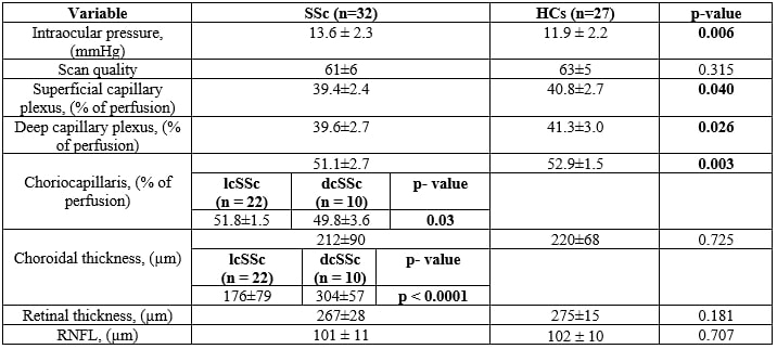
Abbreviations. SSc: systemic sclerosis; HCs: healthy controls; lcSSc: localized cutaneous systemic sclerosis; dcSSc: diffuse cutaneous systemic sclerosis; RNFL= retinal nerve fiber layer.


Disclosures: C. Cutolo: None; E. Hysa: None; A. Cere: None; P. Toma: None; T. Cannavacciuolo: None; C. Toma: None; S. Balito: None; V. Gerli: None; A. Sulli: None; V. Smith: Boehringer Ingelheim, 2, 5, 6, 12, Support for travel, Galapagos, 6, Janssen-Cilag, 1, 2, 5, 6; C. Traverso: None; M. Nicolò: None; M. Cutolo: Amgen, 5, Boehringer Ingelheim, 5, Bristol-Myers Squibb, 5, Lab.Baldacci, 5.

To cite this abstract in AMA style:

Cutolo C, Hysa E, Cere A, Toma P, Cannavacciuolo T, Toma C, Balito S, Gerli V, Sulli A, Smith V, Traverso C, Nicolò M, Cutolo M. Optical Coherence Tomography Angiography in Systemic Sclerosis: Correlations with Morphological and Functional Peripheral Microvascular Status [abstract]. Arthritis Rheumatol. 2023; 75 (suppl 9). https://acrabstracts.org/abstract/optical-coherence-tomography-angiography-in-systemic-sclerosis-correlations-with-morphological-and-functional-peripheral-microvascular-status/. Accessed .
  • Tweet
  • Click to email a link to a friend (Opens in new window) Email
  • Click to print (Opens in new window) Print

« Back to ACR Convergence 2023

ACR Meeting Abstracts - https://acrabstracts.org/abstract/optical-coherence-tomography-angiography-in-systemic-sclerosis-correlations-with-morphological-and-functional-peripheral-microvascular-status/

Advanced Search

Your Favorites

You can save and print a list of your favorite abstracts during your browser session by clicking the “Favorite” button at the bottom of any abstract. View your favorites »

All abstracts accepted to ACR Convergence are under media embargo once the ACR has notified presenters of their abstract’s acceptance. They may be presented at other meetings or published as manuscripts after this time but should not be discussed in non-scholarly venues or outlets. The following embargo policies are strictly enforced by the ACR.

Accepted abstracts are made available to the public online in advance of the meeting and are published in a special online supplement of our scientific journal, Arthritis & Rheumatology. Information contained in those abstracts may not be released until the abstracts appear online. In an exception to the media embargo, academic institutions, private organizations, and companies with products whose value may be influenced by information contained in an abstract may issue a press release to coincide with the availability of an ACR abstract on the ACR website. However, the ACR continues to require that information that goes beyond that contained in the abstract (e.g., discussion of the abstract done as part of editorial news coverage) is under media embargo until 10:00 AM ET on November 14, 2024. Journalists with access to embargoed information cannot release articles or editorial news coverage before this time. Editorial news coverage is considered original articles/videos developed by employed journalists to report facts, commentary, and subject matter expert quotes in a narrative form using a variety of sources (e.g., research, announcements, press releases, events, etc.).

Violation of this policy may result in the abstract being withdrawn from the meeting and other measures deemed appropriate. Authors are responsible for notifying colleagues, institutions, communications firms, and all other stakeholders related to the development or promotion of the abstract about this policy. If you have questions about the ACR abstract embargo policy, please contact ACR abstracts staff at [email protected].

Wiley

  • Online Journal
  • Privacy Policy
  • Permissions Policies
  • Cookie Preferences

© Copyright 2025 American College of Rheumatology