ACR Meeting Abstracts

ACR Meeting Abstracts

  • Meetings
    • ACR Convergence 2024
    • ACR Convergence 2023
    • 2023 ACR/ARP PRSYM
    • ACR Convergence 2022
    • ACR Convergence 2021
    • ACR Convergence 2020
    • 2020 ACR/ARP PRSYM
    • 2019 ACR/ARP Annual Meeting
    • 2018-2009 Meetings
    • Download Abstracts
  • Keyword Index
  • Advanced Search
  • Your Favorites
    • Favorites
    • Login
    • View and print all favorites
    • Clear all your favorites
  • ACR Meetings

Abstract Number: 1185

New Imaging Modality to Evaluate Arthritis in Lupus Based on Frequency-domain Optical Transmission

Anca Askanase1, Fiamma Van Biema 2, Alessandro Marone 2, Youngwan Kim 2, Tommy Chen 2, Teja Kapoor 2, Laura Geraldino 2, Yevgeniya Gartshteyn 3 and Andreas Hielscher 2, 1Columbia University Medical Center, New York, NY, 2Columbia University School of Medicine, New York, NY, 3Columbia University School of Medicine, Glen Rock, NJ

Meeting: 2019 ACR/ARP Annual Meeting

Keywords: Arthritis, Imaging and radiology, Lupus

  • Tweet
  • Click to email a link to a friend (Opens in new window) Email
  • Click to print (Opens in new window) Print
Session Information

Date: Monday, November 11, 2019

Title: Imaging Of Rheumatic Diseases Poster II

Session Type: Poster Session (Monday)

Session Time: 9:00AM-11:00AM

Background/Purpose: 95% of the people with SLE experience joint pain, stiffness and swelling at some time during their illness. It is currently difficult to both diagnose and estimate the severity of lupus arthritis.This studyexplores the clinical utility of frequency-domain optical transmission (FDOT) measurements to distinguish affected finger joints from healthy ones.

Methods: The FDOT system is operated at a frequency of 600MHz. The laser diode produces a 1-mm diameter beam at 670nm light with a power of 8mW. Transmitted light intensity is measured using an ICCD camera operated in homodyne mode.

2

1

For data acquisition, the laser beam is guided to 11 positions, separated by 2 mm from each other, on the top of the finger, scanning along a sagittal plane in the forward direction with a modulation frequency of 600 MHz. PIP II -V of both hands were scanned. The time required to set-up the system and scan a single finger was about 2 min. 10 patients with SLE with active arthritis were evaluated (9 women, mean age 42 ± 9.1 years). All patients had arthritis by physical exam: swelling, tenderness and pain in each finger joint. 4 healthy controls were also examined (3 women, mean age 28.7 ± 4.4 years). A total of 80 fingers from SLE patients and 32 fingers from healthy volunteers were analyzed.

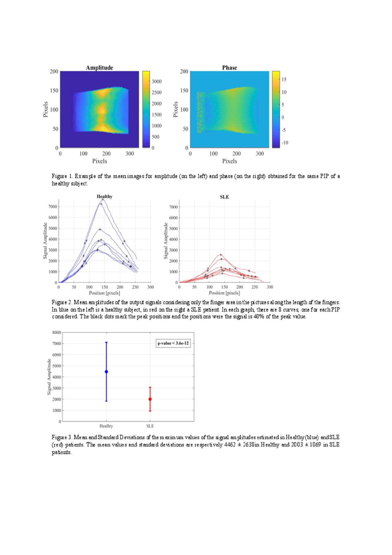
Results: The raw data obtained consisted in 16 pictures for each of the 11 source positions for a total of 176 images per finger. The data was processed using a fast Fourier transformation, obtaining values for the amplitude, phase and the DC components for all source positions. An example of the 2D distribution of the mean values for amplitude and phase for a healthy PIP subject can be seen in Figure 1. From the 2D images, a linear mean value of the amplitudes and phases along the length of the finger was calculated. An example of the amplitude behaviors from healthy joints and SLE joints are shown in Figure 2.

The maximum values of the signal amplitudes in healthy volunteers is considerably higher (4462 ± 2638) than in SLE patients (2003 ± 1069). An ANOVA analysis (using MATLAB embedded function anova1) shows that these differences are statistically significant between healthy and SLE patients (p value < 3.6e-11), as shown in Figure 3.

From the ROC curve analysis, the best cut-off plane holds a sensitivity of 100% and a specificity of 80% to distinguish between joints of Healthy volunteers and SLE patients, with an area under the curve (AUC) of 0.9.

Conclusion: FDOT can differentiate healthy joints from those of SLE patients. We foundstatistically significant differences between the max signal amplitudes of the two groups. The ROC analysis showed high sensitivity and specificity.  Further development of this technology is likely to improve the evaluation of SLE arthritis in clinical trials and practice.


Tables FDOT


Disclosure: A. Askanase, None; F. Van Biema, None; A. Marone, None; Y. Kim, None; T. Chen, None; T. Kapoor, None; L. Geraldino, None; Y. Gartshteyn, None; A. Hielscher, None.

To cite this abstract in AMA style:

Askanase A, Van Biema F, Marone A, Kim Y, Chen T, Kapoor T, Geraldino L, Gartshteyn Y, Hielscher A. New Imaging Modality to Evaluate Arthritis in Lupus Based on Frequency-domain Optical Transmission [abstract]. Arthritis Rheumatol. 2019; 71 (suppl 10). https://acrabstracts.org/abstract/new-imaging-modality-to-evaluate-arthritis-in-lupus-based-on-frequency-domain-optical-transmission/. Accessed .
  • Tweet
  • Click to email a link to a friend (Opens in new window) Email
  • Click to print (Opens in new window) Print

« Back to 2019 ACR/ARP Annual Meeting

ACR Meeting Abstracts - https://acrabstracts.org/abstract/new-imaging-modality-to-evaluate-arthritis-in-lupus-based-on-frequency-domain-optical-transmission/

Advanced Search

Your Favorites

You can save and print a list of your favorite abstracts during your browser session by clicking the “Favorite” button at the bottom of any abstract. View your favorites »

All abstracts accepted to ACR Convergence are under media embargo once the ACR has notified presenters of their abstract’s acceptance. They may be presented at other meetings or published as manuscripts after this time but should not be discussed in non-scholarly venues or outlets. The following embargo policies are strictly enforced by the ACR.

Accepted abstracts are made available to the public online in advance of the meeting and are published in a special online supplement of our scientific journal, Arthritis & Rheumatology. Information contained in those abstracts may not be released until the abstracts appear online. In an exception to the media embargo, academic institutions, private organizations, and companies with products whose value may be influenced by information contained in an abstract may issue a press release to coincide with the availability of an ACR abstract on the ACR website. However, the ACR continues to require that information that goes beyond that contained in the abstract (e.g., discussion of the abstract done as part of editorial news coverage) is under media embargo until 10:00 AM ET on November 14, 2024. Journalists with access to embargoed information cannot release articles or editorial news coverage before this time. Editorial news coverage is considered original articles/videos developed by employed journalists to report facts, commentary, and subject matter expert quotes in a narrative form using a variety of sources (e.g., research, announcements, press releases, events, etc.).

Violation of this policy may result in the abstract being withdrawn from the meeting and other measures deemed appropriate. Authors are responsible for notifying colleagues, institutions, communications firms, and all other stakeholders related to the development or promotion of the abstract about this policy. If you have questions about the ACR abstract embargo policy, please contact ACR abstracts staff at [email protected].

Wiley

  • Online Journal
  • Privacy Policy
  • Permissions Policies
  • Cookie Preferences

© Copyright 2025 American College of Rheumatology