ACR Meeting Abstracts

ACR Meeting Abstracts

  • Meetings
    • ACR Convergence 2024
    • ACR Convergence 2023
    • 2023 ACR/ARP PRSYM
    • ACR Convergence 2022
    • ACR Convergence 2021
    • ACR Convergence 2020
    • 2020 ACR/ARP PRSYM
    • 2019 ACR/ARP Annual Meeting
    • 2018-2009 Meetings
    • Download Abstracts
  • Keyword Index
  • Advanced Search
  • Your Favorites
    • Favorites
    • Login
    • View and print all favorites
    • Clear all your favorites
  • ACR Meetings

Abstract Number: 0242

Neural Networks for Distinguishing Rheumatoid Arthritis from Psoriatic Arthritis by Using Magnetic Resonance Imaging

Lukas Folle1, Sara Bayat2, Arnd Kleyer3, Filippo Fagni2, Lorenz Kapsner4, Maja Schlereth5, Timo Meinderink2, Katharina Breininger5, Koray Tascilar3, Gerhard Kroenke3, Michael Uder6, MIchael Sticherling7, Sebastian Bickelhaupt4, Georg Schett8, Andreas Maier9, Frank Roemer10 and David Simon3, 1Friedrich-Alexander-Universitaet Erlangen Nürnberg, Erlangen, Germany, 2University Hospital Erlangen; Department of Internal Medicine 3 - Rheumatology and Immunology, Erlangen, Germany, 3Department of Internal Medicine 3 – Rheumatology and Immunology, Friedrich-Alexander-University Erlangen-Nürnberg and Universitätsklinikum Erlangen, 91054 Erlangen, Germany; Deutsches Zentrum Immuntherapie, Friedrich-Alexander-UniversityErlangen-Nürnberg and Universitätsklinikum Erlangen, Erlangen, Germany, 4University Hospital Erlangen; Institute of Radiology, Erlangen, Germany, 5Friedrich-Alexander Universität Erlangen-Nürnberg, Artificial Intelligence in Medical Imaging, Erlangen, Germany, 6University Hospital Erlangen; Lehrstuhl für Diagnostische Radiologie, Erlangen, Germany, 7University Hospital Erlangen; Institute of Dermatology, Erlangen, Germany, 8Universitätsklinikum Erlangen, Erlangen, Germany, 9Friedrich-Alexander-Universität Erlangen-Nürnberg, Lehrstuhl für Mustererkennung, Erlangen, Germany, 10Friedrich-Alexander University Erlangen Nürnberg, Erlangen, Germany

Meeting: ACR Convergence 2022

Keywords: hand, informatics, Magnetic resonance imaging (MRI), rheumatoid arthritis

  • Tweet
  • Click to email a link to a friend (Opens in new window) Email
  • Click to print (Opens in new window) Print
Session Information

Date: Saturday, November 12, 2022

Title: RA – Diagnosis, Manifestations, and Outcomes Poster I

Session Type: Poster Session A

Session Time: 1:00PM-3:00PM

Background/Purpose: To evaluate (i) whether neural networks can distinguish seropositive rheumatoid arthritis (RA+), seronegative RA (RA-), and psoriatic arthritis (PsA) using MRI data based on the structural inflammation patterns.

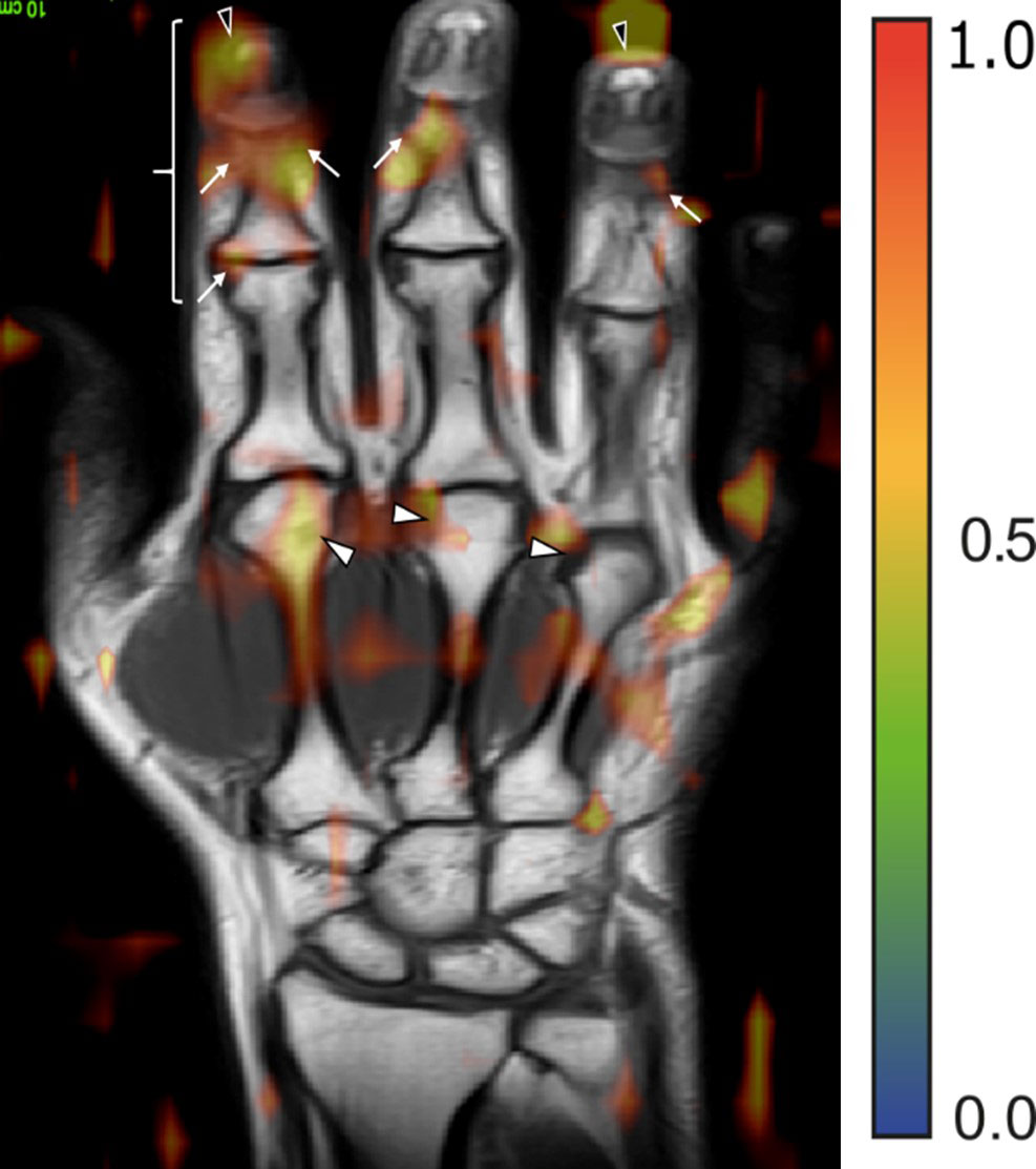
Methods: Neural networks based on the ResNet [1] architecture were trained to distinguish (i) RA+ vs. PsA, (ii) RA- vs. PsA and (iii) RA+ vs. RA- using hand MRI data. MR sequences consisted of five different sequences (T1 coronal, T2 coronal, T1 coronal and axial fat suppressed contrast-enhanced (CE), and T2 fat suppressed axial). Two neural network trainings were performed, where (i) the neural network had access only to MR data and (ii) then to MR and clinical and demographic data (Figure 1). Using a specialized visualization technique for the neural networks (occlusion) the regions most important for the network were recorded and correlated to the respective anatomical regions by an experienced rheumatologist [2,3].

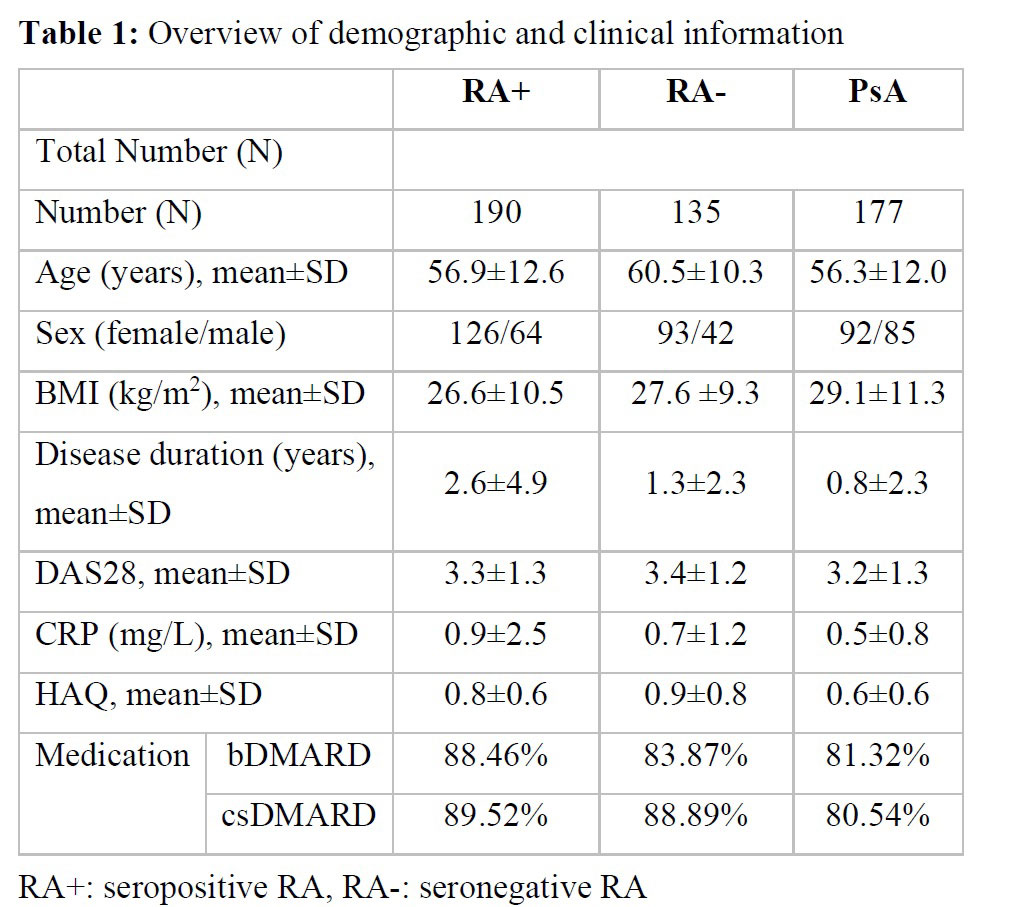
Results: The dataset consisted of MRI scans from 502 patients (135 RA-, 190 RA+, 177 PsA) (Table 1). Differentiation between disease entities as measured by the AUROC was 75% (SD 3%) for RA+ vs PsA, 74% (SD 8%) for RA- vs PsA, and 67% (6%) for RA+ vs RA-. All MR sequences were important for neural network performance, but without contrast-enhanced sequences, the difference in performance was marginal. Adding demographic and clinical parameters to MR dada did not improve the performance of the neural network significantly. Interestingly, the regions important for the network were regions, which also had clinical relevance (Figure 2).

Conclusion: Neural networks can help distinguish between different forms of inflammatory arthritis based on MRI inflammatory patterns, apparently focusing on regions of clinical relevance.

References [1] Kensho Hara, Hirokatsu Kataoka, and Yutaka Satoh 2018. Can Spatiotemporal 3D CNNs Retrace the History of 2D CNNs and ImageNet? In Proceedings of the IEEE Conference on Computer Vision and Pattern Recognition (CVPR) (pp. 6546–6555).
[2] Østergaard M, McQueen F, Wiell C, et al. The OMERACT Psoriatic Arthritis Magnetic Resonance Imaging Scoring System (PsAMRIS): Definitions of key pathologies, suggested MRI sequences, and preliminary scoring system for PsA hands. J Rheumatol 2009;36:1816–24. doi:10.3899/jrheum.090352
[3] Østergaard M, Peterfy C, Conaghan P, et al. OMERACT Rheumatoid Arthritis Magnetic Resonance Imaging Studies. Core set of MRI acquisitions, joint pathology definitions, and the OMERACT RA-MRI scoring system. J Rheumatol 2003;30:1385–6.

Supporting image 1

Table 1: Overview of demographic and clinical information

Supporting image 2

Figure 1: Neural network combining all MR sequences along with the optional additional clinical and demographical data. The prediction for a single case is formed by averaging the prediction of all sequences and the clinical data.

Supporting image 3

Figure 2: MR image with the overlay of the attention map generated by the neural network for a patient affected by PsA. Arrows mark regions of pathological joint changes (soft tissue inflammation and enthesitis).


Disclosures: L. Folle, None; S. Bayat, None; A. Kleyer, None; F. Fagni, None; L. Kapsner, None; M. Schlereth, None; T. Meinderink, None; K. Breininger, None; K. Tascilar, Gılead, AbbVie/Abbott, UCB, Eli Lilly; G. Kroenke, None; M. Uder, None; M. Sticherling, None; S. Bickelhaupt, None; G. Schett, None; A. Maier, None; F. Roemer, Boston Imaging Core Lab (BICL) LLC., Grünenthal, Calibr; D. Simon, None.

To cite this abstract in AMA style:

Folle L, Bayat S, Kleyer A, Fagni F, Kapsner L, Schlereth M, Meinderink T, Breininger K, Tascilar K, Kroenke G, Uder M, Sticherling M, Bickelhaupt S, Schett G, Maier A, Roemer F, Simon D. Neural Networks for Distinguishing Rheumatoid Arthritis from Psoriatic Arthritis by Using Magnetic Resonance Imaging [abstract]. Arthritis Rheumatol. 2022; 74 (suppl 9). https://acrabstracts.org/abstract/neural-networks-for-distinguishing-rheumatoid-arthritis-from-psoriatic-arthritis-by-using-magnetic-resonance-imaging/. Accessed .
  • Tweet
  • Click to email a link to a friend (Opens in new window) Email
  • Click to print (Opens in new window) Print

« Back to ACR Convergence 2022

ACR Meeting Abstracts - https://acrabstracts.org/abstract/neural-networks-for-distinguishing-rheumatoid-arthritis-from-psoriatic-arthritis-by-using-magnetic-resonance-imaging/

Advanced Search

Your Favorites

You can save and print a list of your favorite abstracts during your browser session by clicking the “Favorite” button at the bottom of any abstract. View your favorites »

All abstracts accepted to ACR Convergence are under media embargo once the ACR has notified presenters of their abstract’s acceptance. They may be presented at other meetings or published as manuscripts after this time but should not be discussed in non-scholarly venues or outlets. The following embargo policies are strictly enforced by the ACR.

Accepted abstracts are made available to the public online in advance of the meeting and are published in a special online supplement of our scientific journal, Arthritis & Rheumatology. Information contained in those abstracts may not be released until the abstracts appear online. In an exception to the media embargo, academic institutions, private organizations, and companies with products whose value may be influenced by information contained in an abstract may issue a press release to coincide with the availability of an ACR abstract on the ACR website. However, the ACR continues to require that information that goes beyond that contained in the abstract (e.g., discussion of the abstract done as part of editorial news coverage) is under media embargo until 10:00 AM ET on November 14, 2024. Journalists with access to embargoed information cannot release articles or editorial news coverage before this time. Editorial news coverage is considered original articles/videos developed by employed journalists to report facts, commentary, and subject matter expert quotes in a narrative form using a variety of sources (e.g., research, announcements, press releases, events, etc.).

Violation of this policy may result in the abstract being withdrawn from the meeting and other measures deemed appropriate. Authors are responsible for notifying colleagues, institutions, communications firms, and all other stakeholders related to the development or promotion of the abstract about this policy. If you have questions about the ACR abstract embargo policy, please contact ACR abstracts staff at [email protected].

Wiley

  • Online Journal
  • Privacy Policy
  • Permissions Policies
  • Cookie Preferences

© Copyright 2025 American College of Rheumatology