
Session Information
Session Type: ACR Poster Session A
Session Time: 9:00AM-11:00AM
Background/Purpose: Near infrared (NIR) imaging studies of subdermal indocyanine green (ICG) in murine models of inflammatory arthritis established the important contribution of lymphatic vessel (LV) function in joint homeostasis and flare. However, the role of lymphatic vessel function and altered joint drainage in rheumatoid arthritis (RA) flare is unknown.
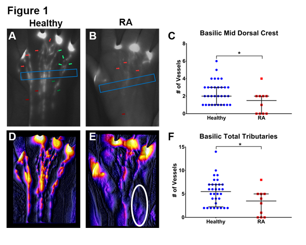
Methods: The web spaces in both hands of 7 healthy controls (Ctl) and 2 subjects in RA flare were injected with 0.1ml of 100μM ICG on 2-4 separate occasions and the NIR fluorescence of the dorsal aspect imaged. Two independent graders counted the total number of fluorescent LVs crossing the mid-dorsal aspect of the hand, and the number of LVs associated with the basilic and cephalic veins and their tributaries. Differences between Ctl vs RA were evaluated using Fisher’s Exact Test, pooling left and right hand observations across all visits (n=32 Ctl, n=10 RA). The frequency of lymphatic contractions of these LVs were determined from graphs of region of interest intensity across time to calculate contractions per minute; differences evaluated using a Wilcoxon Rank Test.
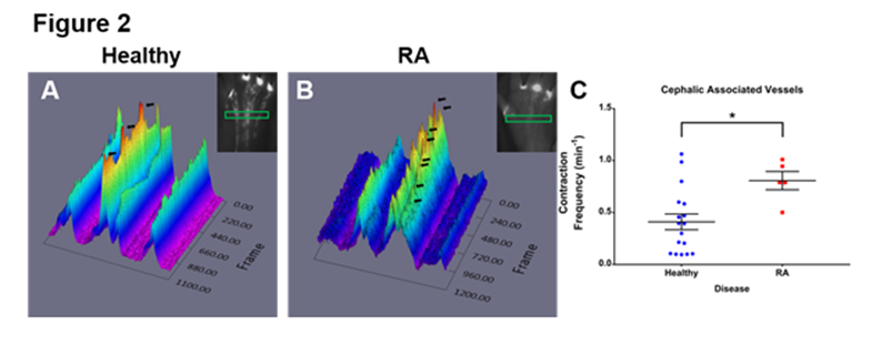
Results: Representative raw images of Ctl and RA hands are presented in Fig 1A-B demonstrating the Mid-Dorsal region (Blue Box), Basilic (Dark Green Arrows) and Cephalic (Dark Red) associated clusters and their tributaries (Basilic = Light Green, Cephalic = Light Red). The number of basilic associated LVs crossing the mid dorsal region and the feeding tributaries were decreased in RA subjects compared to controls (Fig 1C and F, p<0.05). Contour plots generated by stabilizing and averaging all images during the 10min session show clear anatomic and bulk flow differences (Fig 1 D-E). Note the lack of Basilic LV filled with ICG in RA (circled region in E). Representative 3D plots of the ROI analysis are shown in Fig 2A and B with scored contractions (Black Arrows) revealing a significant increase in contraction frequency in the cephalic associated LVs in RA subjects (Fig 2C, p<0.05).
Conclusion: For the first time, we have characterized the lymphatic vessel anatomy of the hands of healthy subjects and RA subject using NIR ICG imaging. Furthermore, RA patients in flare show a reduced number of fluorescent LVs in the hands compared to Ctl. A decline in the number of LVs removing inflammatory cells and molecules from the joints and compensatory redirection of flow is likely to play a significant role in RA flare. Further studies to confirm these initial findings are underway.
To cite this abstract in AMA style:
Bell R, Lieberman A, Wood R, Bell C, Rahimi H, Schwarz E, Ritchlin CT. Near Infrared Indocyanine Green Imaging Reveals Diminished Flow in Basilic Associated Lymphatic Vessels in the Hands of Rheumatoid Arthritis Patients during Flare [abstract]. Arthritis Rheumatol. 2017; 69 (suppl 10). https://acrabstracts.org/abstract/near-infrared-indocyanine-green-imaging-reveals-diminished-flow-in-basilic-associated-lymphatic-vessels-in-the-hands-of-rheumatoid-arthritis-patients-during-flare/. Accessed .« Back to 2017 ACR/ARHP Annual Meeting
ACR Meeting Abstracts - https://acrabstracts.org/abstract/near-infrared-indocyanine-green-imaging-reveals-diminished-flow-in-basilic-associated-lymphatic-vessels-in-the-hands-of-rheumatoid-arthritis-patients-during-flare/