ACR Meeting Abstracts

ACR Meeting Abstracts

  • Meetings
    • ACR Convergence 2024
    • ACR Convergence 2023
    • 2023 ACR/ARP PRSYM
    • ACR Convergence 2022
    • ACR Convergence 2021
    • ACR Convergence 2020
    • 2020 ACR/ARP PRSYM
    • 2019 ACR/ARP Annual Meeting
    • 2018-2009 Meetings
    • Download Abstracts
  • Keyword Index
  • Advanced Search
  • Your Favorites
    • Favorites
    • Login
    • View and print all favorites
    • Clear all your favorites
  • ACR Meetings

Abstract Number: 1177

Near Infrared Indocyanine Green Imaging Reveals Altered Anatomy and Diminished Function in Lymphatic Vessels in the Hands of Rheumatoid Arthritis Patients During Flare

Richard Bell1, Homaira Rahimi 2, Alicia Lieberman 2, Ronald Wood 2, Edward Schwarz 3 and Christopher Ritchlin 4, 1University of Rochester Medical Center, Rochester, NY, 2University of Rochester Medical Center, Rochester, 3University of Rochester, Rochester, NY, 4Division of Allergy, Immunology and Rheumatology, Center for Musculoskeletal Research, University of Rochester School of Medicine and Dentistry, Rochester, NY, USA, Rochester, NY

Meeting: 2019 ACR/ARP Annual Meeting

Keywords: Imaging, lymph, outcome measures and Flare, Rheumatoid arthritis (RA)

  • Tweet
  • Email
  • Print
Session Information

Date: Monday, November 11, 2019

Title: Imaging Of Rheumatic Diseases Poster II

Session Type: Poster Session (Monday)

Session Time: 9:00AM-11:00AM

Background/Purpose: Near infrared (NIR) imaging of indocyanine green (ICG) in rheumatoid arthritis (RA) models identified abnormal lymphatic vessel (LV) function, which can be quantified as ICG clearance from the injection site via longitudinal (days) NIR imaging.  As the role of LV function in RA is unknown, we hypothesized that ICG clearance from hand web spaces and efferent LV contraction frequency of RA patients experiencing flare is significantly decreased compared to normal healthy volunteers. We also assessed the characteristics of filled LVs to examine if structural differences are present in RA lymphatics and healthy controls.

Methods: The web spaces of both hands of 12 healthy controls (CTL) and 7 subjects with RA flare (ACR 2010 RA criteria) were injected with 0.1ml of 100μM ICG on 2-4 separate occasions, and the hands underwent NIR imaging. To measure clearance of ICG from the web spaces, 7 CTL and 7 RA subjects were reimaged 7-9 or 13-15 days after the first injections, and the remaining NIR fluorescence was measured via region of interest (ROI) analysis (Paired t-test). Controlling for the number of days between subjects, the change in intensity between the initial and second visit was divided by the number of days between visits (t-test). Lymphatic contractions for LVs were determined from graphs of ROI intensity across time to calculate contractions per minute, and assessed via Wilcoxon Rank Test. To assess the branching structure of the vessels, manual segmentation of the lymphatic network and subsequent spatial mapping was performed (t-test). Then two independent graders quantified the total number of bifurcations of the LVs. Median values for each hand across all visits and graders were used to test for differences (Wilcoxon Rank-Sum Test).

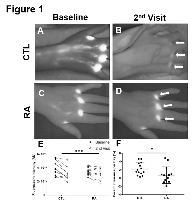
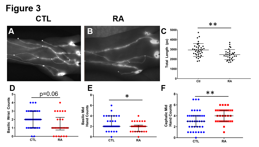
Results: Representative NIR images of CTL and RA hands at baseline, and at the second visit, demonstrate the dramatic retention of ICG at the injections sites (White Arrows) in RA subjects (Fig 1 A-D). Statistical analysis revealed a decrease in ICG clearance in the RA subjects vs. CTLs (Fig 1 E, ***p< 0.001). When controlling for days between the initial visit and second visit the relationship persisted (Fig 1F, *p< 0.05). Interestingly, no difference in contraction frequency was observed between CTLs and RA subjects (Fig 2A-D). However, there was significantly decreased total length in the spatial structure of the lymphatic network (Fig 3A-C, *p< 0.05).  This decreased length was primarily attributed to a lack of basilic associated ICG filled vessels in RA subjects (Fig 3D-F, *p< 0.05).

Conclusion: Imaging outcomes of LV function in mice have demonstrated diminished clearance of lymph from inflamed joints during arthritic progression. Herein, we show a significant reduction in ICG clearance and altered anatomic structure in RA subjects’ hands during flare. The accumulation and retention of inflammatory cells and molecules in RA joints as a result of diminished lymphatic clearance may be a critical factor in the initiation and persistence of synovitis. This clinical pilot demonstrates the feasibility of quantifying LV function, and warrants formal investigation in clinical trials.

Figure 1. Impaired Lymphatic Clearance of ICG from the Hand in RA Subjects. ICG intensity was measured in left and right hands of 7 healthy control -CTL, n=14- and 5 RA subjects -n=14- after injection at the initial visit -Baseline- and at the second visit 7-15 days later -2nd Visit-. Representative NIR images of a control subject at baseline -A- and at the second visit -B- show considerable clearance of the ICG at the injection sites -White Arrows-, while in RA subjects -C, D- did not clear the ICG. When this was quantified, RA subjects had significantly less clearance -E, paired t-test,**p<0.001-. Due to the variable time period between baseline and subjects second visit, the percent clearance per day is plotted for each interval -F, t-test, *p<0.05-.

Figure 2. Lymphatic contraction frequency is not different between healthy controls and RA subjects. Lymphatic contraction frequency was calculated via ROI analysis of 10 minutes observing the dorsal aspect of the hand. An ROI was placed over the basilic and cephalic associated vessels at the wrist. These data for each vessel were either averaged or summed to generate average basilic -A- and cephalic -B- contraction frequency; or total basilic -C- and cephalic -D- contraction frequency. There were no statistical difference between the groups for any of these outcomes.

Figure 3. RA subjects have significantly less lymphatic vessels in their hands compared to healthy controls. Videos and still images were used to manually segment the lymphatic vessel network in the dorsal aspects of the hands. Spatial graphs were then generated to map to the lymphatic network. Representative stills of CTL -A- and RA -B- hands with overlaid spatial graphs show significant variation between the two. Accordingly, RA subjects have significantly less total graph length is calculated -C, t-test, M± SD, **p<0.01-. To investigate where anatomically these lymphatic network differences, we manually scored how many vessels were present at the wrist, mid hand and near the injection sites. Interestingly, we found significantly fewer vessels on the lateral side of the hand -basilic associated vessels, D and E- while there was an increase in cephalic sided vessels -F, Wilcoxon Sum Rank Test, Median and IQR, *p<0.05, **0.001-.


Disclosure: R. Bell, None; H. Rahimi, None; A. Lieberman, None; R. Wood, None; E. Schwarz, None; C. Ritchlin, AbbVie, 2, 5, 9, Amgen, 2, 5, BMS, 5, Janssen, 5, Janssen Research & Development, LLC, 2, Lilly, 5, Novartis, 5, Pfizer, 2, Pfizer Inc, 5, UCB, 2, 5.

To cite this abstract in AMA style:

Bell R, Rahimi H, Lieberman A, Wood R, Schwarz E, Ritchlin C. Near Infrared Indocyanine Green Imaging Reveals Altered Anatomy and Diminished Function in Lymphatic Vessels in the Hands of Rheumatoid Arthritis Patients During Flare [abstract]. Arthritis Rheumatol. 2019; 71 (suppl 10). https://acrabstracts.org/abstract/near-infrared-indocyanine-green-imaging-reveals-altered-anatomy-and-diminished-function-in-lymphatic-vessels-in-the-hands-of-rheumatoid-arthritis-patients-during-flare/. Accessed .
  • Tweet
  • Email
  • Print

« Back to 2019 ACR/ARP Annual Meeting

ACR Meeting Abstracts - https://acrabstracts.org/abstract/near-infrared-indocyanine-green-imaging-reveals-altered-anatomy-and-diminished-function-in-lymphatic-vessels-in-the-hands-of-rheumatoid-arthritis-patients-during-flare/

Advanced Search

Your Favorites

You can save and print a list of your favorite abstracts during your browser session by clicking the “Favorite” button at the bottom of any abstract. View your favorites »

All abstracts accepted to ACR Convergence are under media embargo once the ACR has notified presenters of their abstract’s acceptance. They may be presented at other meetings or published as manuscripts after this time but should not be discussed in non-scholarly venues or outlets. The following embargo policies are strictly enforced by the ACR.

Accepted abstracts are made available to the public online in advance of the meeting and are published in a special online supplement of our scientific journal, Arthritis & Rheumatology. Information contained in those abstracts may not be released until the abstracts appear online. In an exception to the media embargo, academic institutions, private organizations, and companies with products whose value may be influenced by information contained in an abstract may issue a press release to coincide with the availability of an ACR abstract on the ACR website. However, the ACR continues to require that information that goes beyond that contained in the abstract (e.g., discussion of the abstract done as part of editorial news coverage) is under media embargo until 10:00 AM ET on November 14, 2024. Journalists with access to embargoed information cannot release articles or editorial news coverage before this time. Editorial news coverage is considered original articles/videos developed by employed journalists to report facts, commentary, and subject matter expert quotes in a narrative form using a variety of sources (e.g., research, announcements, press releases, events, etc.).

Violation of this policy may result in the abstract being withdrawn from the meeting and other measures deemed appropriate. Authors are responsible for notifying colleagues, institutions, communications firms, and all other stakeholders related to the development or promotion of the abstract about this policy. If you have questions about the ACR abstract embargo policy, please contact ACR abstracts staff at [email protected].

Wiley

  • Online Journal
  • Privacy Policy
  • Permissions Policies
  • Cookie Preferences

© Copyright 2025 American College of Rheumatology