Session Information
Date: Sunday, November 12, 2023
Title: (0283–0307) Muscle Biology, Myositis & Myopathies – Basic & Clinical Science Poster I
Session Type: Poster Session A
Session Time: 9:00AM-11:00AM
Background/Purpose: Muscular involvement develops as the initial manifestation of small-sized vessel vasculitis (SV) and medium-sized vessel vasculitis (MV).The musculoskeletal lesion has been found as a crucial biopsy site including the histology of SV and MV unless another suitable organ for a biopsy can be found. However, immunopathological features, including the degree of myofiber damage, in the histology of vasculitic myopathy (VM) have been still unknown, while pathological assessment is an ideal procedure for the definite diagnosis of vasculitis. We elucidated the immunopathological features of skeletal muscle in VM by comparing the immunohistochemical (IHC) findings of skeletal muscle in patients with idiopathic inflammatory myositis (IIM).
Methods: The biopsied skeletal muscle tissues from 15 patients with VM, including antineutrophil cytoplasmic antibody-associated vasculitis and polyarteritis nodosa, and 15 with IIM, including polymyositis and immune-mediated necrotizing myopathy, were used in this study. IHC staining of skeletal muscle was performed as follows: CD56/neural cell adhesion molecule (NCAM) which detects myofiber damage and regeneration, major histocompatibility complex (MHC)-class I, C5b-9/membrane attack complex (MAC), and CD31 which is an endothelial cell marker. The vascularity score was defined as the total number of CD31-expressing blood vessels.
The counts and calculations of IHC stained samples and the vascularity scores were performed in the 10 different high-power fields.
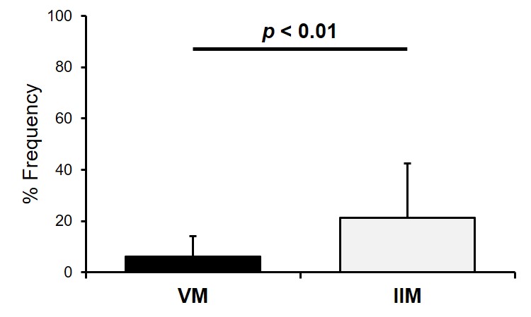
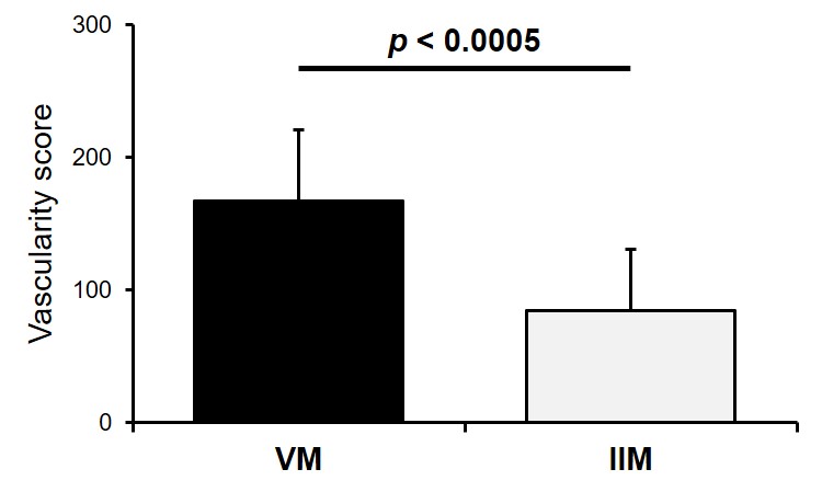
Results: The frequency of NCAM-expressing myofibers was significantly lower in patients with VM than in those with IIM (p < 0.01) (Fig. 1). In addition, the frequency of NCAM-expressing myofibers was positively correlated with serum aldolase levels (p < 0.01). The frequency of MHC class I-expressing myofibers was significantly lower in patients with VM than in those with IIM (p < 0.005). A lower number of patients with MV had MAC-expressing myofibers on sarcolemma without intracellular staining than those with IIM (33% vs. 80%, p < 0.05).Meanwhile, the frequency of patients, who had MAC-expressing capillaries in endomysium areas, was not significantly different between VM and IIM (73% vs. 93%). Significantly higher vascularity scores in the endomysium areas were demonstrated in patients with MV than in those with IIM (p < 0.0005) (Fig. 2).
Conclusion: Mild myofiber damage, based on the result of less involving NCAM-expressing myofiber, was demonstrated in patients with VM than in those with IIM. Our results suggest that complement component deposits on vessel walls and hypervascularity in the endomysium areas may be immunopathological features of VM.
To cite this abstract in AMA style:
Nomura S, Shimojima Y, Ichikawa T, Miyazaki D, Kishida D, Sekijima Y. Myopathy Related to Small-to-medium-sized Vessel Vasculitis: Immunopathological Characteristics [abstract]. Arthritis Rheumatol. 2023; 75 (suppl 9). https://acrabstracts.org/abstract/myopathy-related-to-small-to-medium-sized-vessel-vasculitis-immunopathological-characteristics/. Accessed .« Back to ACR Convergence 2023
ACR Meeting Abstracts - https://acrabstracts.org/abstract/myopathy-related-to-small-to-medium-sized-vessel-vasculitis-immunopathological-characteristics/


