ACR Meeting Abstracts

ACR Meeting Abstracts

  • Meetings
    • ACR Convergence 2024
    • ACR Convergence 2023
    • 2023 ACR/ARP PRSYM
    • ACR Convergence 2022
    • ACR Convergence 2021
    • ACR Convergence 2020
    • 2020 ACR/ARP PRSYM
    • 2019 ACR/ARP Annual Meeting
    • 2018-2009 Meetings
    • Download Abstracts
  • Keyword Index
  • Advanced Search
  • Your Favorites
    • Favorites
    • Login
    • View and print all favorites
    • Clear all your favorites
  • ACR Meetings

Abstract Number: 1621

Maxillofacial MRI Augments Detection of Non-CNS Abnormalities in Pediatric Craniofacial Scleroderma

Daniel Glaser1, Amy Metry2, Elena DeRosas2, Subramanian Subramanian2 and Kathryn Torok3, 1Yale University School of Medicine, New Haven, CT, 2University of Pittsburgh, Pittsburgh, PA, 3University of Pittsburgh, University of Pittsburgh Medical Center, Pittsburgh, PA

Meeting: ACR Convergence 2023

Keywords: Magnetic resonance imaging (MRI), Pediatric rheumatology, Scleroderma

  • Tweet
  • Click to email a link to a friend (Opens in new window) Email
  • Click to print (Opens in new window) Print
Session Information

Date: Monday, November 13, 2023

Title: Abstracts: Pediatric Rheumatology – Clinical II: Connective Tissue Disease

Session Type: Abstract Session

Session Time: 2:00PM-3:30PM

Background/Purpose: Craniofacial scleroderma is a rare subtype of linear scleroderma affecting the head and neck. Up to 40% of patients experience extracutaneous manifestations (ECMs), but radiographic characterization of non-CNS involvement is not well described. Existing clinical assessment rubrics prioritize cutaneous changes, likely underestimating the extent of disease-related manifestations. We sought to categorize and quantify the incidence of non-CNS ECMs detected on maxillofacial MRI in a prospective cohort of patients with craniofacial scleroderma. We hypothesize that maxillofacial MRI would identify facial involvement beyond standard clinical examination.

Methods: We performed retrospective review of 120 patients with Craniofacial Scleroderma enrolled in the prospective National Registry of Childhood Onset Scleroderma (IRB #19080297). Inclusion criteria included baseline maxillofacial MRI with concurrent clinical 3D image.

Laterality of visible facial involvement (left, right, midline) and superior to inferior facial lesion distribution (top, middle, bottom thirds) were recorded from review of 3-dimensional digital facial images. Top third was defined as the area between horizontal lines drawn through trichion and glabella, middle third between glabella and subnasale, and bottom third between subnasale and mention. Evaluation of bone, soft tissue, fat, glands, and muscles were categorized as atrophy, edema, or abnormal enhancement.

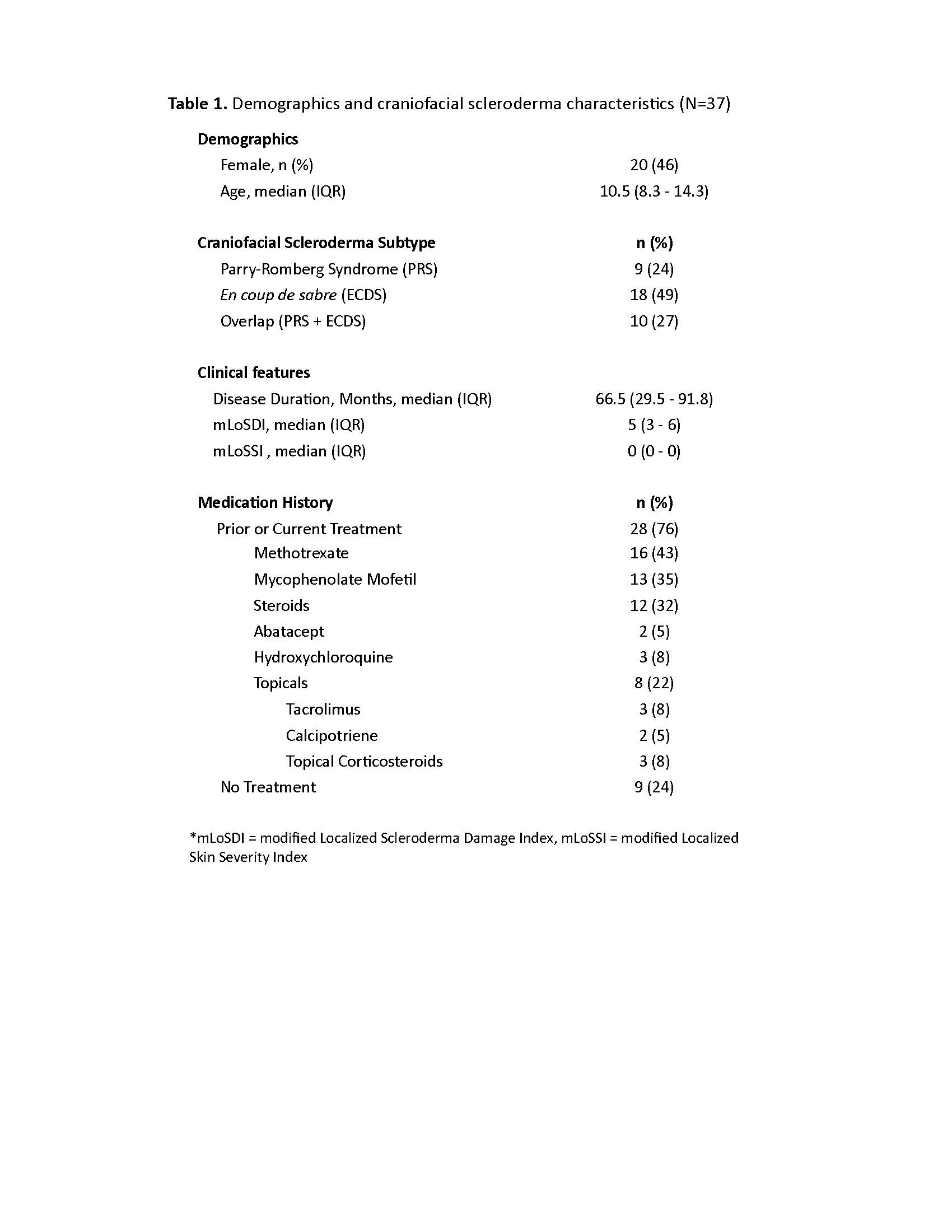
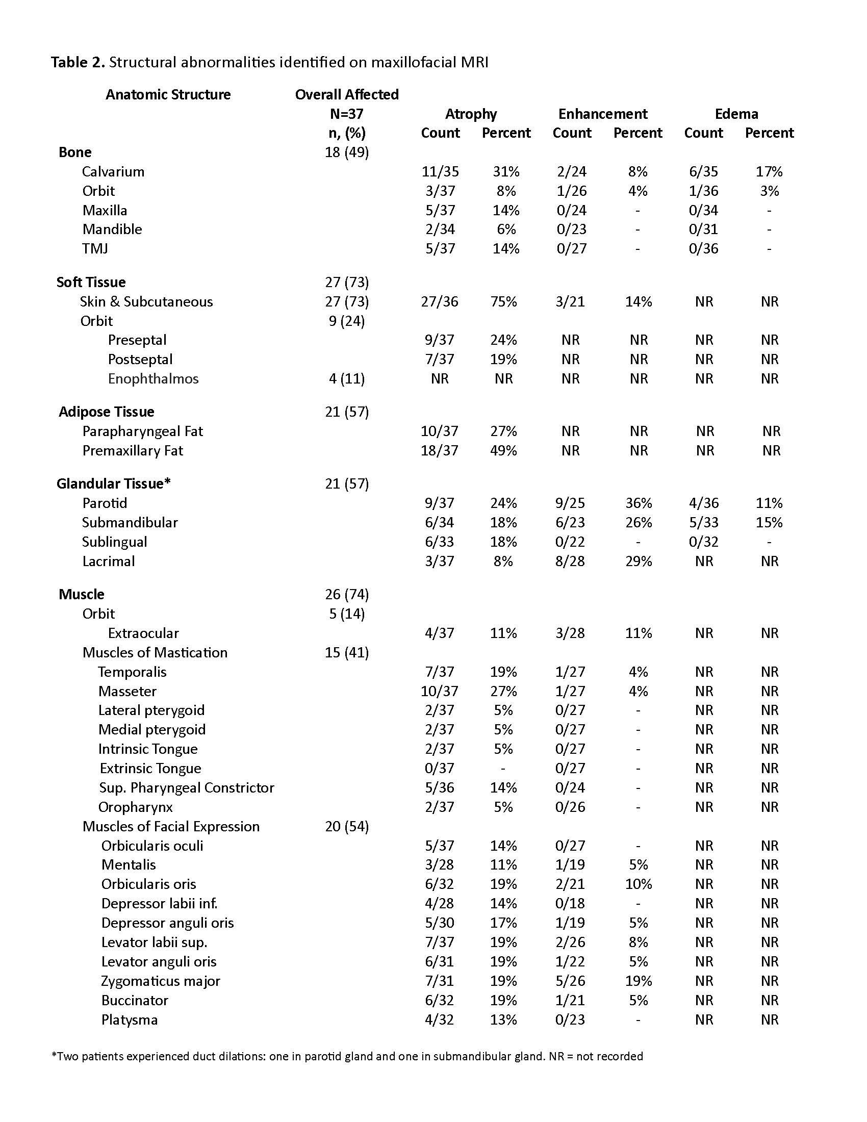
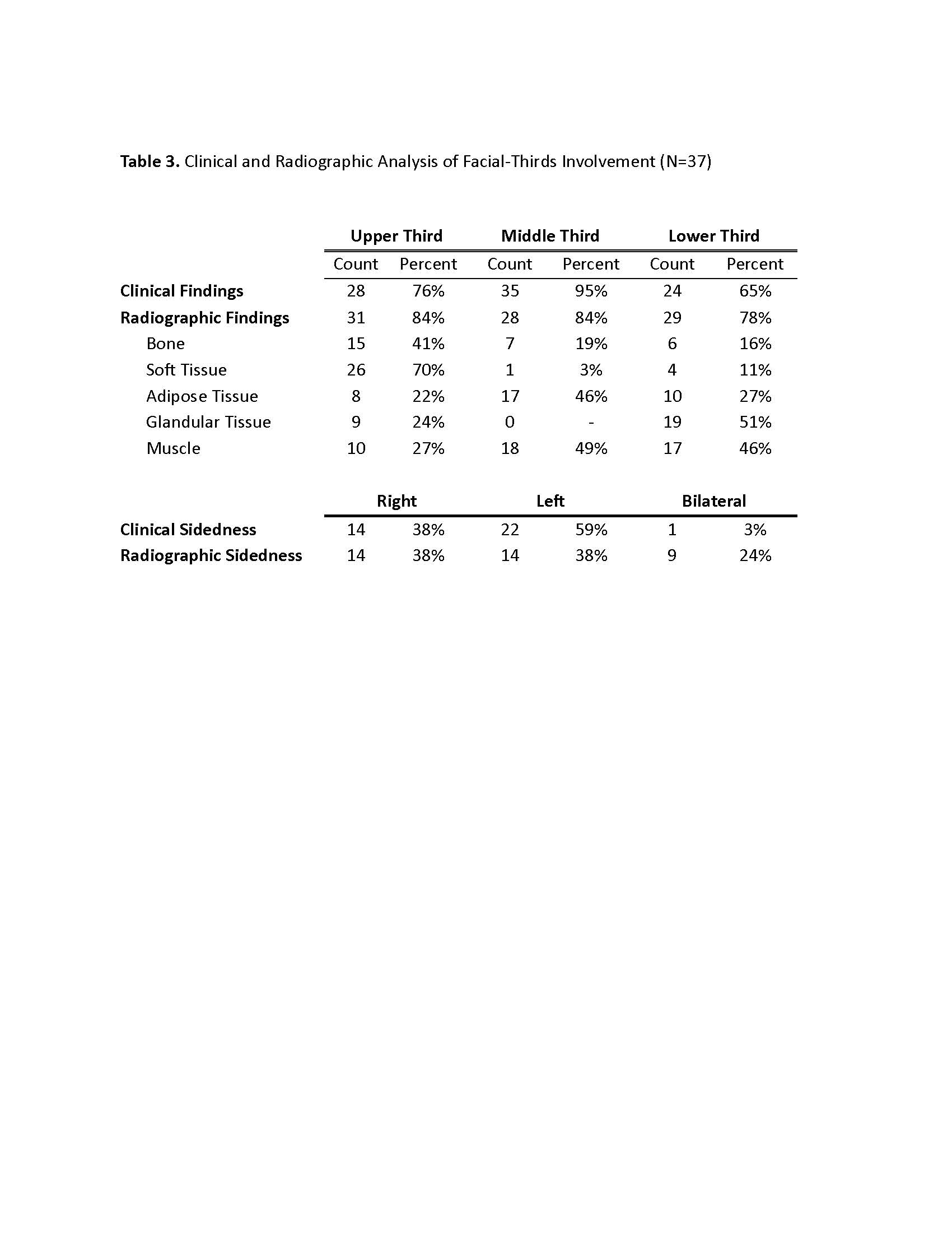
Results: Thirty-seven patients met imaging criteria for inclusion, (age range 4-20 years, median age 10.5 years) (Table 1). Overall, structural abnormalities were frequently detected on maxillofacial MRI (bone=49%, soft tissue=73%, adipose=57%, gland=57% and muscle=74%) (Table 2). Atrophy was the most common finding, particularly in the skin/subcutis (75%) and adipose tissues (57%). Enhancement was frequently observed in glandular tissues (parotid 36%, lacrimal 29%) as well as in the muscles of facial expression. MRI demonstrated increased frequency of bilateral involvement compared to clinical assessment (24% vs 3%). Radiographic changes were more frequently seen compared to clinical abnormalities in the upper-third (84% vs 76%) and lower-third (78% vs 65%) (Table 3). Frequencies of radiographic abnormalities differed across facial thirds, with soft tissue changes most frequent in the upper third (70%), muscle changes in the middle third (49%), and glandular changes in the lower third (51%).

Conclusion: Our study demonstrated an overall high incidence of facial bone, soft tissue, glandular, and muscle involvement in craniofacial scleroderma. Clinical examination and radiographic review frequently demonstrated discordant results. Therefore, we recommended baseline maxillofacial MRI in addition to brain MRI to better delineate the full extent of disease involvement in craniofacial scleroderma patients. Future work should assess evolution of these findings over time, response to immunomodulatory treatment, and development of multi-modal assessment rubrics.

Supporting image 1

Table 1. Demographics and craniofacial scleroderma characteristics (N=37)

Supporting image 2

Table 2. Structural abnormalities identified on maxillofacial MRI

Supporting image 3

Table 3. Clinical and radiographic analysis of facial-thirds involvement (N=37)


Disclosures: D. Glaser: None; A. Metry: None; E. DeRosas: None; S. Subramanian: None; K. Torok: None.

To cite this abstract in AMA style:

Glaser D, Metry A, DeRosas E, Subramanian S, Torok K. Maxillofacial MRI Augments Detection of Non-CNS Abnormalities in Pediatric Craniofacial Scleroderma [abstract]. Arthritis Rheumatol. 2023; 75 (suppl 9). https://acrabstracts.org/abstract/maxillofacial-mri-augments-detection-of-non-cns-abnormalities-in-pediatric-craniofacial-scleroderma/. Accessed .
  • Tweet
  • Click to email a link to a friend (Opens in new window) Email
  • Click to print (Opens in new window) Print

« Back to ACR Convergence 2023

ACR Meeting Abstracts - https://acrabstracts.org/abstract/maxillofacial-mri-augments-detection-of-non-cns-abnormalities-in-pediatric-craniofacial-scleroderma/

Advanced Search

Your Favorites

You can save and print a list of your favorite abstracts during your browser session by clicking the “Favorite” button at the bottom of any abstract. View your favorites »

All abstracts accepted to ACR Convergence are under media embargo once the ACR has notified presenters of their abstract’s acceptance. They may be presented at other meetings or published as manuscripts after this time but should not be discussed in non-scholarly venues or outlets. The following embargo policies are strictly enforced by the ACR.

Accepted abstracts are made available to the public online in advance of the meeting and are published in a special online supplement of our scientific journal, Arthritis & Rheumatology. Information contained in those abstracts may not be released until the abstracts appear online. In an exception to the media embargo, academic institutions, private organizations, and companies with products whose value may be influenced by information contained in an abstract may issue a press release to coincide with the availability of an ACR abstract on the ACR website. However, the ACR continues to require that information that goes beyond that contained in the abstract (e.g., discussion of the abstract done as part of editorial news coverage) is under media embargo until 10:00 AM ET on November 14, 2024. Journalists with access to embargoed information cannot release articles or editorial news coverage before this time. Editorial news coverage is considered original articles/videos developed by employed journalists to report facts, commentary, and subject matter expert quotes in a narrative form using a variety of sources (e.g., research, announcements, press releases, events, etc.).

Violation of this policy may result in the abstract being withdrawn from the meeting and other measures deemed appropriate. Authors are responsible for notifying colleagues, institutions, communications firms, and all other stakeholders related to the development or promotion of the abstract about this policy. If you have questions about the ACR abstract embargo policy, please contact ACR abstracts staff at [email protected].

Wiley

  • Online Journal
  • Privacy Policy
  • Permissions Policies
  • Cookie Preferences

© Copyright 2025 American College of Rheumatology