Session Information
Session Type: Poster Session (Monday)
Session Time: 9:00AM-11:00AM
Background/Purpose: The immune cell subsets most altered early in SLE disease course remain unclear. Defining abnormalities in lymphocyte populations in patients with new-onset SLE may reveal pathways that are fundamental drivers of the autoimmune response in SLE. Mass cytometry provides a powerful method to broadly assess immune cell phenotypes in samples from patients and may reveal immune cell subsets that are most prominently altered in SLE patients compared to controls.
Methods: We used two 39-marker mass cytometry panels (a T cell panel and B cell panel) to evaluate CD4+ T cells and B cells in cryopreserved peripheral blood mononuclear cells from patients with new-onset SLE (diagnosis within 6 months, n=10), compared to patients with established SLE (n=15), and non-inflammatory controls (n=14). All SLE patients met the 1997 ACR classification criteria for SLE. New-onset SLE patients were naïve to immunosuppressive therapy; hydroxychloroquine and prednisone ≤ 10mg/day were permitted. Established SLE patients were on a range of treatments. We used FlowSOM to define and quantify metaclusters of memory CD4+ T cells and B cells based on their 39-parameter characterization. We used Student’s t-test to identify significantly altered metaclusters in SLE patients (p< 0.05). Expanded cell populations were confirmed by biaxial gating.
Results: We identified 15 metaclusters (i.e. cell populations) of memory CD4+ T cells by FlowSOM. Of these, 4 T cell metaclusters were significantly increased in new-onset SLE patients compared to healthy controls (Figure 1). Three of these 4 metaclusters were also increased in established SLE patients. Analysis of marker expression on these cell populations revealed that all 4 populations displayed high expression of PD-1 and ICOS. One of the populations also expressed CXCR5, consistent T follicular helper (Tfh) cells, while the other 3 lacked CXCR5, consistent with T peripheral helper (Tph) cells, a CXCR5- B cell-helper T cell population initially identified in RA joints. Biaxial gating confirmed a significant expansion of both Tph cells (2.0-fold, p=0.034) and Tfh cells (2.4-fold, p=0.001) in new-onset SLE patients. In the analysis of B cells by FlowSOM, we identified 3/15 B cell metaclusters significantly expanded in new-onset SLE patients (Figure 2). Cells in all 3 of these metaclusters showed a CD11c+ Tbet+ CD21- phenotype consistent with age-associated B cells (ABCs). The 3 metaclusters were distinguished by expression of distinct immunoglobulin isotypes representing IgM+, IgG+, and IgA+ ABCs. Across all patient groups, the frequency of Tph cells was positively correlated with the frequency of ABCs (r=0.34,p=0.036), including both IgG+ and IgM+ ABCs, and with the frequency of Tfh cells (r=0.44,p=0.005). Tph cells, but not Tfh cells, also showed a negative association with serum complement C3 levels (r=-0.51,p=0.017), a marker of disease activity in SLE.
Conclusion: Broad immunophenotyping analyses highlight marked expansion of Tph cells, Tfh cells, and ABCs are prominent features of the immune dysregulation in patients with new-onset, immunosuppressant-naïve SLE. These cells may be involved in the early phases of the pathologic autoimmune response in SLE.
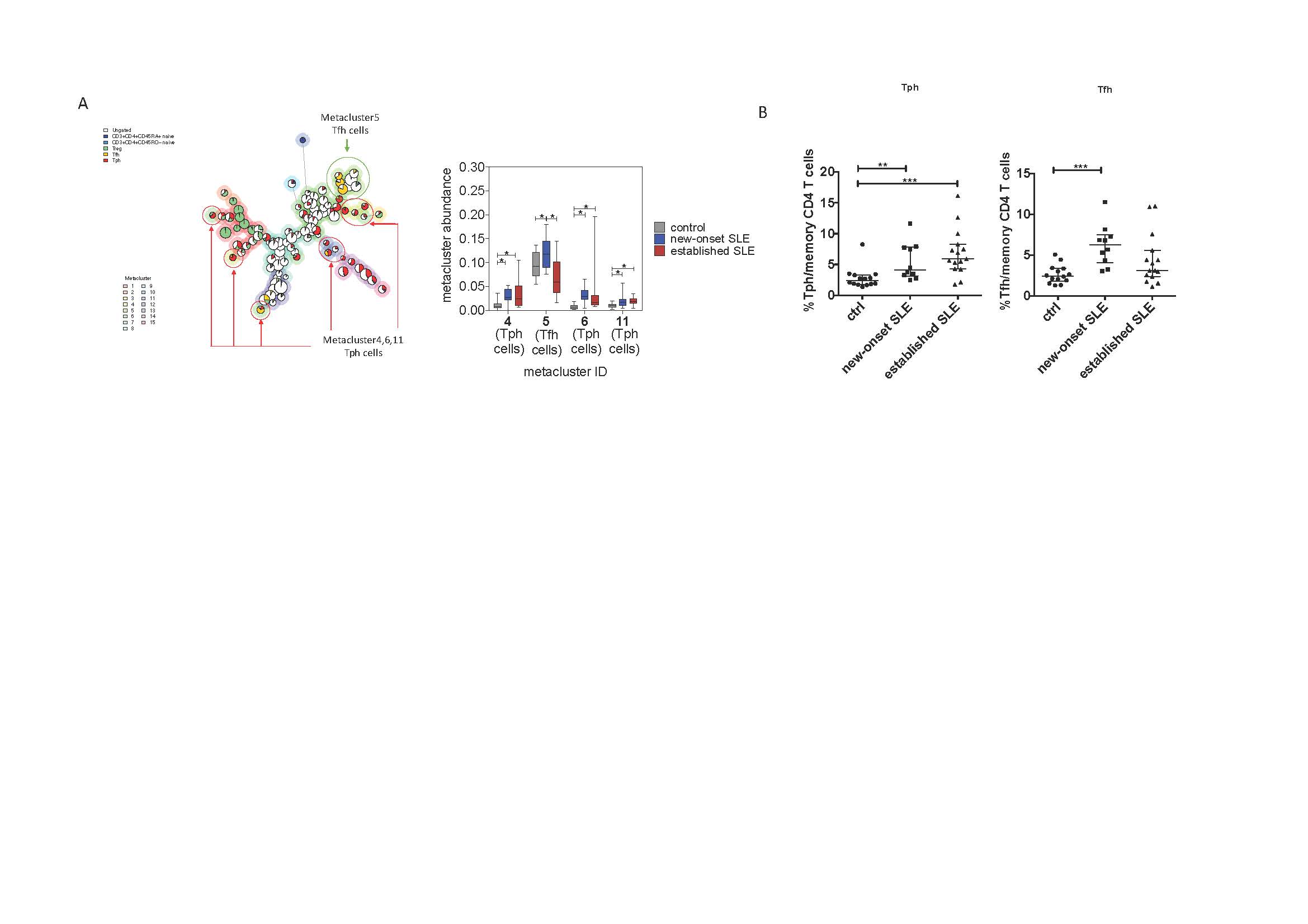
ACR_Figure1
A. FlowSOM analysis of CD45RO+ CD4+ T cells shows an increased abundance of T cell metaclusters 4,5,6,11. Metaclusters 4,6,11 are PD-1hi CXCR5- cells. Metacluster 5 is PD-1hi CXCR5+ cells. *Student’s t-test p<0.05.
B. Quantification of Tph cells -PD-1hi CXCR5– and Tfh cells -PD-1hi CXCR5+- cells by biaxial gating. * p<0.05, ** p<0.01, *** p<0.001 by Mann-Whitney U test.
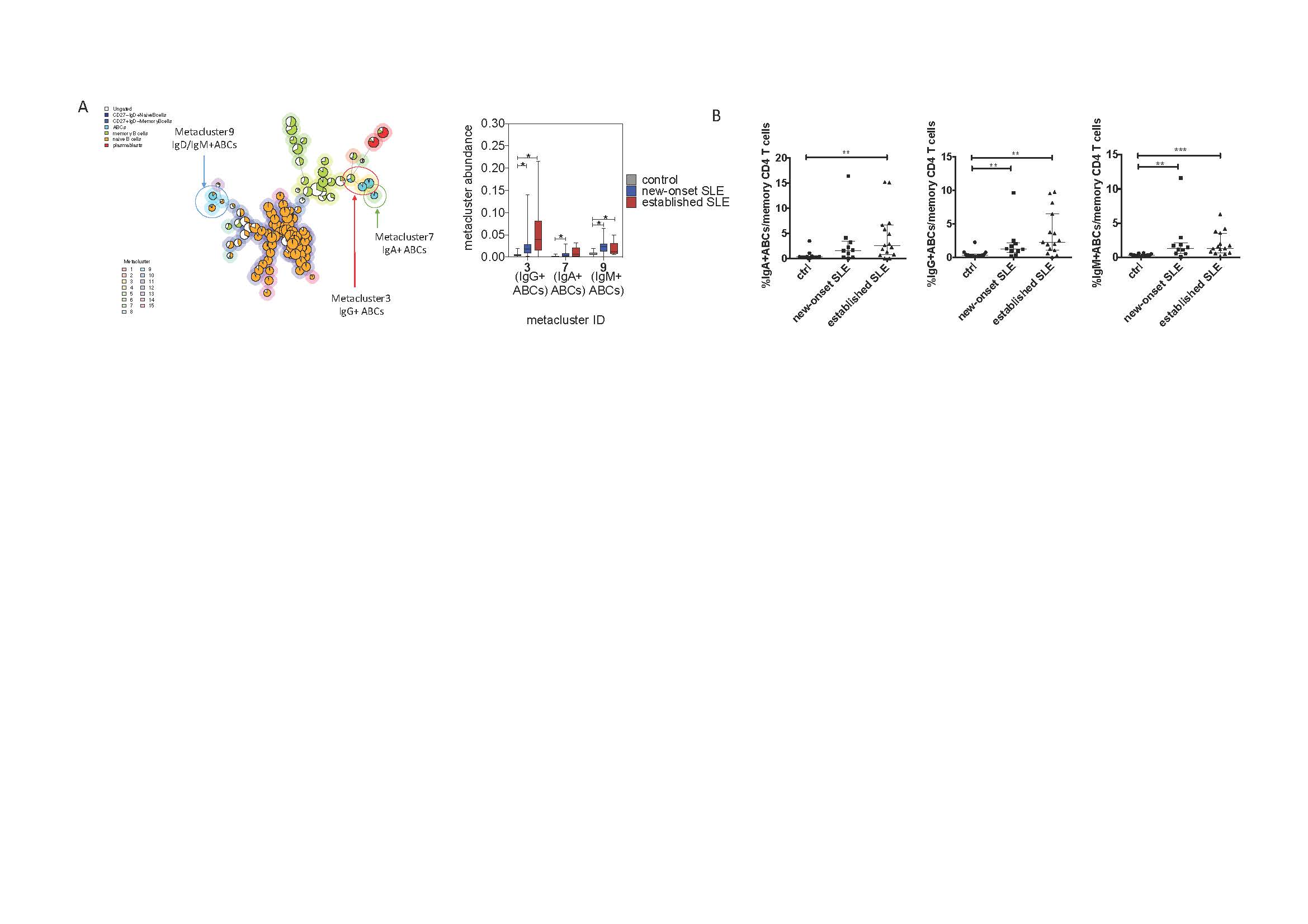
ACR_Figure2
A. FlowSOM analysis of total B cells shows an increased abundance of B cell metaclusters 3,7 and 9. Metacluster 3 is IgG+ ABCs. Metacluster 7 is IgA+ ABCs. Metacluster 9 is IgM+ ABCs. *Student’s t-test p<0.05.
B. Quantification of IgG+, IgA+, and IgM+ ABCs by biaxial gating. * p<0.05, ** p<0.01, *** p<0.001 by by Mann-Whitney U test.
To cite this abstract in AMA style:
Cao Y, Alves S, Sinnette C, Speyer C, Keras G, Qu Y, Keegan J, Lederer J, Rao D, Costenbader K. Mass Cytometric Immunophenotyping Highlights a Dysregulated T cell-B Cell Axis in Patients with New-onset Lupus [abstract]. Arthritis Rheumatol. 2019; 71 (suppl 10). https://acrabstracts.org/abstract/mass-cytometric-immunophenotyping-highlights-a-dysregulated-t-cell-b-cell-axis-in-patients-with-new-onset-lupus/. Accessed .« Back to 2019 ACR/ARP Annual Meeting
ACR Meeting Abstracts - https://acrabstracts.org/abstract/mass-cytometric-immunophenotyping-highlights-a-dysregulated-t-cell-b-cell-axis-in-patients-with-new-onset-lupus/