Session Information
Date: Tuesday, October 28, 2025
Title: (1780–1808) Osteoarthritis & Joint Biology – Basic Science Poster
Session Type: Poster Session C
Session Time: 10:30AM-12:30PM
Background/Purpose: Knee joints are densely innervated by sensory neurons, predominantly nociceptors. Knowledge about innervation of healthy human synovium and the changes that accompany osteoarthritis (OA) is limited. Very few studies described innervation changes in select areas of synovium collected at total knee arthroplasty (1-3). Here, we sought to perform a comprehensive analysis of the sensory innervation of the synovium of healthy human knees.
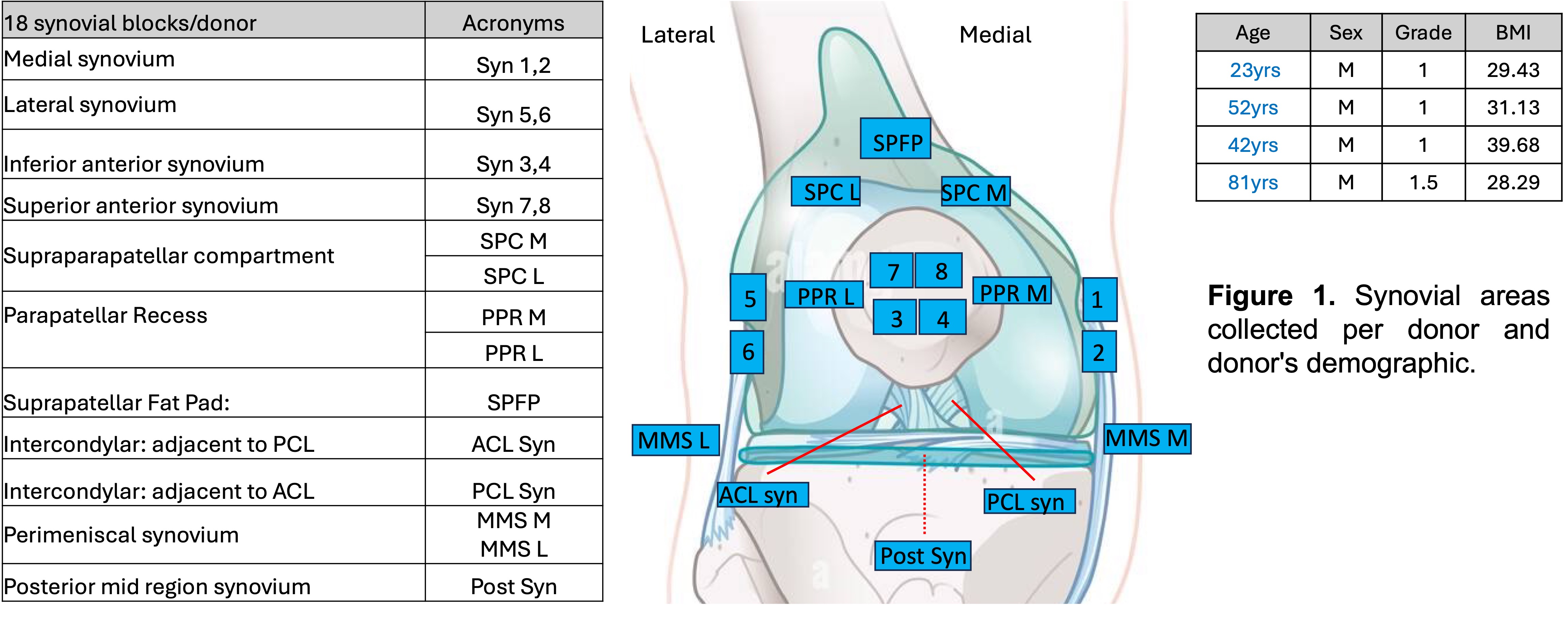
Methods: Leveraging the availability of non-OA postmortem knee samples from human donors of different ages, we collected synovia from 4 males, ages 23, 42, 52 and 81, all from knees where cartilage was scored as grade 1-1.5 on the Modified Outerbridge classification. For each donor, a total of 18 tissue blocks were collected from 18 distinct synovial locations in the knee Fig. 1. These areas are medial (area 1,2) and lateral (area 5,6) femoral gutters, medial and lateral perimeniscal synovium (MMS M, MMS L) and anterior synovium (areas 3,4,7,8), the synovium covering the anterior cruciate ligament (ACL) and posterior cruciate ligament (PCL), medial and lateral suprapatellar compartments (SPC M, SPC L), medial and lateral parapatellar recess (PPR M, PPR L), posterior synovium (Post syn), and suprapatellar fat pad (SPFP). Tissues were formalin fixed, paraffin embedded and shipped from Scripps to Rush. Five-mm sections were stained with hematoxylin and eosin (H&E) and sent to the Hospital for Special Surgery for synovial scoring. Adjacent sections were used for immunohistochemical staining with the pan-neuronal marker, PGP9.5 (Abcam ab27053, 1:200), endothelial cell markers (CD31, Abcam Ab28364, 1:50), calcitonin gene related peptide (CGRP, immunostar 24112,1:500) or isotype control (Rabbit IgG, Abcam Ab172730, 1mg/ml). Proteinase K was used for antigen retrieval. Sections were incubated with primary antibody at 4°C overnight, stained with biotin-streptavidin/HRP and DAB chromogen, and counterstained with methyl green. PGP9.5+ and CGRP+ signal was quantified in adipose and fibrous areas of synovium using Qupath by 2 blinded observers.
Results: In all 18 regions, PGP9.5+ innervation was denser in the adipose rich areas compared to the fibrous areas. PGP9.5+ nerve signal was abundant in the synovium of medial and lateral femoral gutters, medial and lateral suprapatellar compartment, anterior synovium and synovium of the suprapatellar fat pad. PGP9.5+ nerves were more abundant in the medial synovium of the 81yr-old compared to younger donors Fig. 2. PGP9.5 innervation of SPFP and anterior and lateral perimeniscal synovium declined with age but increased in the lateral femoral gutters. Further characterization of the synovial samples, including CD31 staining for blood vessels is ongoing.
Conclusion: PGP9.5+ nerve fibers were abundant in medial, lateral and anterior synovium of non-OA male donors aged 23 to 81, but a marked heterogeneity was observed across the 18 regions. Adipose rich synovium showed more innervation compared to fibrous synovium. These findings are the foundation for future studies on describing the innervation changes in the OA synovium. Saito T et al., Clin Orthop. 2000.Eitner et al., OAC. 2013.Eitner A et al., Front Endocrinol. 2024.
.jpg)
To cite this abstract in AMA style:
obeidat a, Li J, Fullam S, Carter R, Olmer M, Mehta B, Otero M, Ramirez D, Miller R, Orange D, Lotz M, Miller R, Malfait A. Mapping The Synovial Innervation Of The Human Knee Joint [abstract]. Arthritis Rheumatol. 2025; 77 (suppl 9). https://acrabstracts.org/abstract/mapping-the-synovial-innervation-of-the-human-knee-joint/. Accessed .« Back to ACR Convergence 2025
ACR Meeting Abstracts - https://acrabstracts.org/abstract/mapping-the-synovial-innervation-of-the-human-knee-joint/