Session Information
Date: Sunday, November 5, 2017
Title: Spondyloarthropathies and Psoriatic Arthritis – Clinical Aspects and Treatment Poster I
Session Type: ACR Poster Session A
Session Time: 9:00AM-11:00AM
Background/Purpose : A newly developed scoring method for low dose CT (ldCT) of the whole spine, CT Syndesmophyte Score (CTSS), has shown good inter-reader reliability and sensitivity to detect changes in bone formation over 2 years in Ankylosing Spondylitis (AS) patients1. Next step in validation is the comparison of assessment of bone formation on ldCT with conventional radiographs (CR).
Methods : Patients from the Sensitive Imaging in Ankylosing Spondylitis cohort were analysed. Inclusion criteria: mNY-criteria+, ³1 syndesmophyte on cervical/lumbar spine CR and _1 inflammatory lesions on MRI-spine. Patients had baseline and two-year CR of the lateral cervical and lumbar spine and whole spine ldCT (approximately 4mSV). Two readers independently assessed CR and ldCT in separate sessions. Images were paired per patient, blinded to time order, patient information, and results of the other imaging technique. CR was assessed using mSASSS. On ldCT, syndesmophytes were scored in coronal and sagittal planes, assessing eight ÔquadrantsÕ per vertebral unit (VU). Scores for syndesmophytes according to the CTSS were: absent=0, <50% of intervertebral disc height (IVDH)=1, _50% of IVDH=2 or IVDH bridging=3. Formation of new syndesmophytes (CR score 0/1¨2/3, CTSS 0¨1/2/3) and growth of existing syndesmophytes (CR score 2¨3, CTSS 1¨2/3, or 2ˆ3) and the combination of new or growing syndesmophytes was calculated per corner/quadrant. Consensus about each outcome was defined by agreement of readers on the same corner/quadrant. The number of syndesmophytes on CR and CT was compared by level. Level 1: upper four quadrants of a VU on ldCT and upper corner on CR. Level 2: lower four quadrants on ldCT and lower corner on CR. Data of CR and ldCT was compared per reader and for the consensus score.
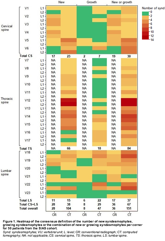
Results : Fifty patients (mean age 50 years; 84% male; 86% HLA-B27+) were included in the analysis. In all comparisons, ldCT detected more patients with progression (table 1). This is especially apparent in case of growth and for cut-offs of a higher number of syndesmophytes per patient. E.g. using the consensus score, 30% of the patients showed bony proliferation (new or growing syndesmophytes) at ³3 sites on ldCT compared with only 6% on CR. ldCT detected more syndesmophytes in all sections of the spine, with most syndesmophytes occurring in the thoracic spine (figure 1).
Conclusion : Whole spine ldCT is more sensitive in assessing the formation and growth of syndesmophytes than CR, which is limited to cervical and lumbar spine and is a promising method of assessment for clinical research of AS.
1. de Bruin F et al. A&R 2016; 68 (suppl 10).
|
Table 1. Comparison of CR and ldCT per reader and consensus* for the formation or growth of syndesmophytes in 50 patients with ankylosing spondylitis. |
|||||||||
|
|
Reader 1 |
Reader 2 |
Consensus |
||||||
|
|
CR n(%) |
ldCT n(%) |
CR n(%) |
ldCT n(%) |
CR n(%) |
ldCT n(%) |
|||
|
New syndesmophytes |
|||||||||
|
³1 |
27 (54) |
43 (86) |
30 (60) |
44 (88) |
19 (38) |
21 (42) |
|||
|
³2 |
14 (28) |
38 (76) |
14 (28) |
41 (82) |
7 (14) |
15 (30) |
|||
|
³3 |
6 (12) |
32 (64) |
8 (16) |
30 (60) |
2 (4) |
10 (20) |
|||
|
Growth of syndesmophytes |
|||||||||
|
³1 |
10 (20) |
35 (70) |
7 (14) |
32 (64) |
3 (6) |
16 (32) |
|||
|
³2 |
8 (16) |
36 (52) |
6 (12) |
27 (54) |
3 (6) |
11 (22) |
|||
|
³3 |
2 (4) |
23 (46) |
4 (8) |
18 (36) |
1 (2) |
6 (12) |
|||
|
New syndesmophytes or growth of syndesmophytes |
|||||||||
|
³1 |
28 (56) |
45 (90) |
33 (66) |
48 (96) |
21 (42) |
25 (50) |
|||
|
³2 |
18 (36) |
42 (82) |
19 (38) |
44 (88) |
9 (18) |
20 (40) |
|||
|
³3 |
12 (24) |
36 (72) |
12 (24) |
38 (76) |
3 (6) |
15 (30) |
|||
|
Results are presented as the number (%) of patients with ³1, ³2 and ³3 newly formed syndesmophytes and syndesmophytes that grew, as well as for the combination of new formation or growth. *Both readers agree about the formation or growth of a syndesmophyte at the same vertebral corner. CR: conventional radiography, ldCT: low-dose computed tomography. |
|||||||||
To cite this abstract in AMA style:
de Koning A, de Bruin F, van den Berg R, Ramiro S, Baraliakos X, Braun J, van Gaalen F, Reijnierse M, van der Heijde D. Low Dose Computed Tomography Detects More Progression of Bone Formation in Comparison to Conventional Radiography in Patients with Ankylosing Spondylitis [abstract]. Arthritis Rheumatol. 2017; 69 (suppl 10). https://acrabstracts.org/abstract/low-dose-computed-tomography-detects-more-progression-of-bone-formation-in-comparison-to-conventional-radiography-in-patients-with-ankylosing-spondylitis/. Accessed .« Back to 2017 ACR/ARHP Annual Meeting
ACR Meeting Abstracts - https://acrabstracts.org/abstract/low-dose-computed-tomography-detects-more-progression-of-bone-formation-in-comparison-to-conventional-radiography-in-patients-with-ankylosing-spondylitis/