ACR Meeting Abstracts

ACR Meeting Abstracts

  • Meetings
    • ACR Convergence 2024
    • ACR Convergence 2023
    • 2023 ACR/ARP PRSYM
    • ACR Convergence 2022
    • ACR Convergence 2021
    • ACR Convergence 2020
    • 2020 ACR/ARP PRSYM
    • 2019 ACR/ARP Annual Meeting
    • 2018-2009 Meetings
    • Download Abstracts
  • Keyword Index
  • Advanced Search
  • Your Favorites
    • Favorites
    • Login
    • View and print all favorites
    • Clear all your favorites
  • ACR Meetings

Abstract Number: 0971

Ligand-Receptor Analysis Reveals Myeloid-Mediated Pro-Inflammatory Responses and Loss of Mesenchymal Signaling Driven by TNFR1 in Experimental Pulmonary Hypertension

Gaochan Wang1, Stacey Duemmel2 and Benjamin Korman3, 1University of Rochester School of Medicine and Dentistry, Rochester, NY, 2URMC, UNIVERSITY OF ROCHESTER, Rochester, NY, 3University of Rochester, Rochester, NY

Meeting: ACR Convergence 2024

Keywords: Animal Model, Scleroderma

  • Tweet
  • Email
  • Print
Session Information

Date: Sunday, November 17, 2024

Title: Systemic Sclerosis & Related Disorders – Basic Science Poster I

Session Type: Poster Session B

Session Time: 10:30AM-12:30PM

Background/Purpose: Pulmonary hypertension (PH) is a severe, progressive disorder characterized by elevated pulmonary artery pressures, right ventricular hypertrophy, and increased mortality which is a severe comorbidity of systemic sclerosis and other connective tissue diseases. We previously demonstrated that TNF-transgenic (TNF-Tg) mice develop a severe PH phenotype and that TNFR1 deficiency reverses this pathology. To explore the roles of cellular interactions contributing to PH in this model, we assessed single-cell RNA expression in TNF-Tg mice crossed with mice lacking TNFR1 or TNFR2.  We found dramatic changes in cell populations between groups, and sought to investigate the cellular communication among immune, endothelial, and stromal populations in the lung.

Methods: TNF-Tg mice were crossed with mice deficient in either TNFR1(TNFR1KO) or TNFR2 (TNFR2KO). Lungs from wild type, TNF-Tg, TNF-Tg /TNFR1KO and TNF-Tg /TNFR2KO mice (n = 5–6) were harvested at 4.5 months of age for histological analysis and single cell RNA sequencing (scRNAseq). The scRNAseq data was analyzed in Seurat v.4.9. Differential expression and pathway analysis were performed and visualized using Seurat and enrichR. Intercellular communication was investigated using NicheNet.

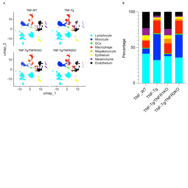
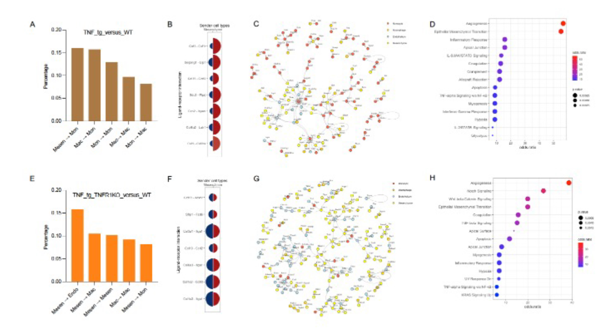
Results: Cells were clustered based on their expression profiles and cell types were annotated based on established cell markers from LungMap (Fig 1A). We observed dramatic expansion of myeloid cells and decreased cellular proportions of endothelial and mesenchymal cell in TNF-Tg and TNF-Tg-TNFR2KO mice as compared to WT and TNF-Tg/TNFR1KO mice (Fig 1B).  We next focused on intercellular interaction among monocyte, macrophage, endothelial, and mesenchymal cells using NicheNet. When we compared signaling between 3 conditions (TNF-Tg, TNF-Tg/TNFR1KO, and TNF-Tg/TNFR2KO) and WT, we identified changes in global cellular communication with a preference for monocyte and macrophage interactions in the TNF-Tg, and TNF-Tg/TNFR2KO conditions (Fig 2A), while TNF-Tg/TNFR1KO mice retained interactions between mesenchymal cells and other cell types (Fig 2E).  In all conditions, angiogenesis and epithelial mesenchymal transition were upregulated in the presence of TNF (Fig 2D/H).  Monocytes and macrophages in the PH conditions mediated inflammatory processes including JAK/Stat, interferon gamma, complement, and TNF/NFkB (Fig 2C/D).  Top ligand-receptor pairs between mesenchymal cells and monocytes include Csf1 with Csf1r, Ccl19 with Ccrl2, and Serping11 with Lrp1 (Fig 2B). Mesenchymal cell-cell interactions in TNF-Tg/TNFR1KO included Notch, Wnt/B-catenin, and TGF-beta pathways which were not seen in the PH conditions (Fig 2G/H).  Top ligand-receptor pairs between mesenchymal and endothelial cells seen in this comparison include Ccl19 with Ackr2, Col1a2 with Cd93, and Sfrp1 with Fzd6.

Conclusion: Our results suggest that TNF-Tg mice develop PH promoted by an increase in myeloid cell infiltrate and corresponding TNF-TNFR1-mediated signaling pathways.  Myeloid cell-cell interactions promote a variety of cellular changes including up-regulation of pro-inflammatory pathways in other cell types, and loss of homeostatic signaling mediated by mesenchymal cells.

Supporting image 1

Figure 1. ScRNA seq demonstrated the changes of cellular composition in TNF-Tg mice. A. UMAP plot of scRNA seq showing 8 distinct cellular clusters. B. Percentage of all cell types revealed the Infiltration of myeloid cells and decrease of endothelial cells and mesenchymal cells found in TNF-Tg and TNF-Tg/TNFR2KO mice. Cell types are indicated by color.

Supporting image 2

Figure 2. Ligand-receptor pairs identified from scRNA seq. A. Top 5 cell-cell interactions was predicted between TNF-Tg and TNF-WT (TNF-Tg_versus_WT). B. Top 7 ligand-receptor pairs from the cellular communication between mesenchymal cells and monocytes. Blue color represents the ligand from mesenchymal cells, red color represents the receptor from monocytes. The shape size represents the percentage of cells expressed ligand or receptor. C. Network analysis of ligand-receptor pairs found only in TNF-Tg_versus_WT. D. Signaling pathway analysis using the unique ligand-receptor pairs found only in TNF-Tg_versus_WT. E. Top 5 cell-cell interactions was predicted between TNF-Tg/TNFR1KO and TNF-WT (TNF-Tg/TNFR1KO_versus_WT). F. Top 7 ligand-receptor pairs from the cellular communication between mesenchymal cells and monocytes. Blue color represents the ligand from mesenchymal cells, red color represents the receptor from monocytes. The shape size represents the percentage of cells expressed ligand or receptor. G. Network analysis of ligand-receptor pairs found only in TNF-Tg/TNFR1KO_versus_WT. H. Signaling pathway analysis using the unique ligand-receptor pairs found only in TNF-Tg/TNFR1KO_versus_WT.


Disclosures: G. Wang: None; S. Duemmel: None; B. Korman: None.

To cite this abstract in AMA style:

Wang G, Duemmel S, Korman B. Ligand-Receptor Analysis Reveals Myeloid-Mediated Pro-Inflammatory Responses and Loss of Mesenchymal Signaling Driven by TNFR1 in Experimental Pulmonary Hypertension [abstract]. Arthritis Rheumatol. 2024; 76 (suppl 9). https://acrabstracts.org/abstract/ligand-receptor-analysis-reveals-myeloid-mediated-pro-inflammatory-responses-and-loss-of-mesenchymal-signaling-driven-by-tnfr1-in-experimental-pulmonary-hypertension/. Accessed .
  • Tweet
  • Email
  • Print

« Back to ACR Convergence 2024

ACR Meeting Abstracts - https://acrabstracts.org/abstract/ligand-receptor-analysis-reveals-myeloid-mediated-pro-inflammatory-responses-and-loss-of-mesenchymal-signaling-driven-by-tnfr1-in-experimental-pulmonary-hypertension/

Advanced Search

Your Favorites

You can save and print a list of your favorite abstracts during your browser session by clicking the “Favorite” button at the bottom of any abstract. View your favorites »

All abstracts accepted to ACR Convergence are under media embargo once the ACR has notified presenters of their abstract’s acceptance. They may be presented at other meetings or published as manuscripts after this time but should not be discussed in non-scholarly venues or outlets. The following embargo policies are strictly enforced by the ACR.

Accepted abstracts are made available to the public online in advance of the meeting and are published in a special online supplement of our scientific journal, Arthritis & Rheumatology. Information contained in those abstracts may not be released until the abstracts appear online. In an exception to the media embargo, academic institutions, private organizations, and companies with products whose value may be influenced by information contained in an abstract may issue a press release to coincide with the availability of an ACR abstract on the ACR website. However, the ACR continues to require that information that goes beyond that contained in the abstract (e.g., discussion of the abstract done as part of editorial news coverage) is under media embargo until 10:00 AM ET on November 14, 2024. Journalists with access to embargoed information cannot release articles or editorial news coverage before this time. Editorial news coverage is considered original articles/videos developed by employed journalists to report facts, commentary, and subject matter expert quotes in a narrative form using a variety of sources (e.g., research, announcements, press releases, events, etc.).

Violation of this policy may result in the abstract being withdrawn from the meeting and other measures deemed appropriate. Authors are responsible for notifying colleagues, institutions, communications firms, and all other stakeholders related to the development or promotion of the abstract about this policy. If you have questions about the ACR abstract embargo policy, please contact ACR abstracts staff at [email protected].

Wiley

  • Online Journal
  • Privacy Policy
  • Permissions Policies
  • Cookie Preferences

© Copyright 2025 American College of Rheumatology