Session Information
Session Type: Poster Session A
Session Time: 9:00AM-11:00AM
Background/Purpose: A knee flexion contracture (FC), present in ~⅓ of patients with knee OA1, is the inability to fully extend the knee2, 3. Having a FC means that anterior and middle regions of the tibial articular surface may experience reduced contact with the femur resulting in altered force transmission, a risk factor for cartilage loss2. The impact of FC on the articular cartilage regions of the tibia has been studied longitudinally in animals2, but not humans. We tested for a quantitative longitudinal association between FC and cartilage loss in the tibia in patients with or at-risk of knee OA using MRI data from the Osteoarthritis Initiative (OAI) cohort. We hypothesized that FC would be associated with cartilage loss in tibial anterior and middle regions, which experience reduced load, but cartilage preservation in the posterior regions, where load is maintained.
Methods: 578 participants with baseline knee extension data and MRI at baseline and year 1 were analyzed. Lack of full knee extension to 0° was considered a FC. Cartilage outcomes were measured using 3T knee MRI coronal views4. Tibial articular cartilage was segmented into medial and lateral compartments, then further divided into anterior, central, and posterior regions4. We looked for associations between the presence of a knee FC, and cartilage thickness or % denuded bone (0mm thickness) over the 1-year period using ANCOVA, controlling for baseline outcomes and relevant covariates.
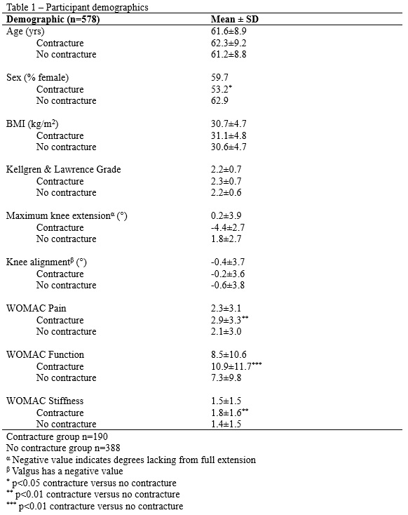
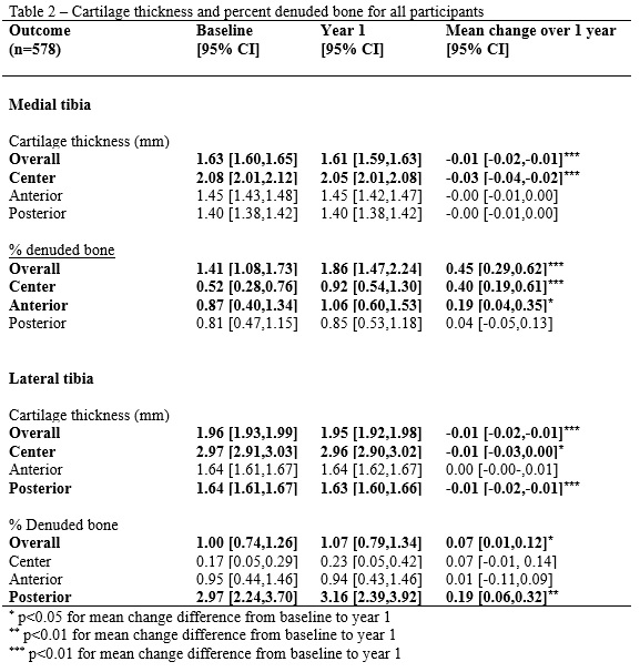
Results: Table 1 shows participant demographics. In the medial compartment, there was reduced cartilage thickness overall and in the center region, with increased % denuded bone overall and in the center and anterior regions. In the lateral compartment, there was reduced cartilage thickness overall and in the center and posterior regions with increased % denuded bone overall and in the posterior region (Table 2).
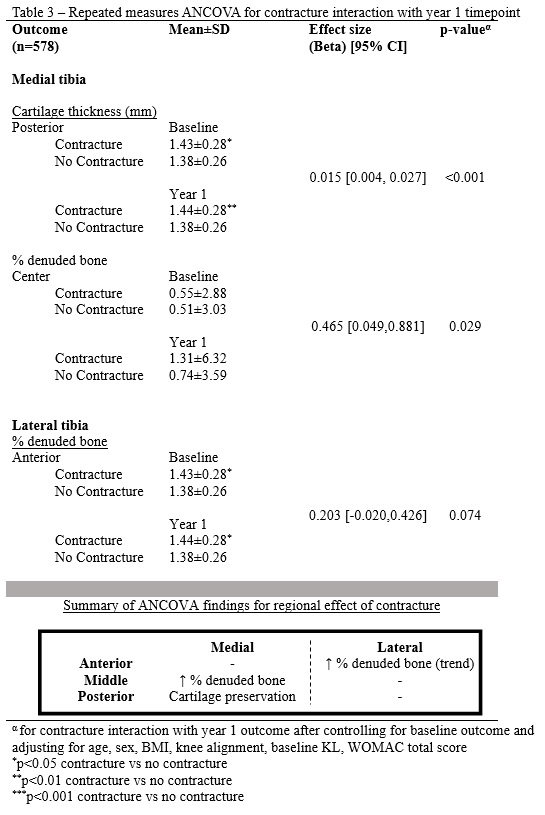
Knee FC was associated with increased % denuded bone in the medial center (β=0.465 [0.049,0.881], p=0.029) and preserved cartilage thickness in the medial posterior (β=0.015 [0.004, 0.027], p< 0.001) regions. There was a trend between FC and increased % denuded bone in the lateral anterior region (β=0.203 [-0.020,0.426], p=0.074; Table 3).
Conclusion: Knee FC was associated with regional cartilage loss in the center region and preservation in the posterior region of the medial tibia, with a trend towards cartilage loss in the lateral anterior region. Together, these findings are consistent with knee FC negatively impacting regions of the tibia articular cartilage with reduced load. Future studies should evaluate the effects of interventions to maintain full knee range of motion on articular cartilage preservation.
References
1. Campbell 2020
2. Watanabe 2020
3. Ritter 2007
4. Wirth 2009
To cite this abstract in AMA style:
Campbell M, Laneuville O, Trudel G. Knee Flexion Contracture Was Associated with Articular Cartilage Loss in the Central Region but Cartilage Preservation in the Posterior Region of the Tibia: Data from the Osteoarthritis Initiative [abstract]. Arthritis Rheumatol. 2023; 75 (suppl 9). https://acrabstracts.org/abstract/knee-flexion-contracture-was-associated-with-articular-cartilage-loss-in-the-central-region-but-cartilage-preservation-in-the-posterior-region-of-the-tibia-data-from-the-osteoarthritis-initiative/. Accessed .« Back to ACR Convergence 2023
ACR Meeting Abstracts - https://acrabstracts.org/abstract/knee-flexion-contracture-was-associated-with-articular-cartilage-loss-in-the-central-region-but-cartilage-preservation-in-the-posterior-region-of-the-tibia-data-from-the-osteoarthritis-initiative/


