ACR Meeting Abstracts

ACR Meeting Abstracts

  • Meetings
    • ACR Convergence 2024
    • ACR Convergence 2023
    • 2023 ACR/ARP PRSYM
    • ACR Convergence 2022
    • ACR Convergence 2021
    • ACR Convergence 2020
    • 2020 ACR/ARP PRSYM
    • 2019 ACR/ARP Annual Meeting
    • 2018-2009 Meetings
    • Download Abstracts
  • Keyword Index
  • Advanced Search
  • Your Favorites
    • Favorites
    • Login
    • View and print all favorites
    • Clear all your favorites
  • ACR Meetings

Abstract Number: 1382

Juvenile Eosinophilic Fasciitis: A Single-Center Cohort

Leigh Stubbs1, Oluwaseun Ogunbona2, Adekunle Adesina1, Sara Anvari1, Emily Beil1, Jamie Lai1, Andrea Ramirez1, Vibha Szafron1, Matthew Ditzler1 and Marietta DeGuzman1, 1Baylor College of Medicine, Houston, TX, 2Houston Methodist Hospital, Houston, TX

Meeting: ACR Convergence 2022

Keywords: Dermatology, Pediatric rheumatology, Scleroderma, Localized

  • Tweet
  • Email
  • Print
Session Information

Date: Sunday, November 13, 2022

Title: Pediatric Rheumatology – Clinical Poster II: Connective Tissue Disease

Session Type: Poster Session C

Session Time: 1:00PM-3:00PM

Background/Purpose: Eosinophilic fasciitis (EF) is a rare fibrosing disease. Since described in 1975, less than 30 pediatric cases have been reported. EF presents with painful swelling and progressive skin induration causing peau d’ orange appearance and the groove sign. Laboratory findings include elevated inflammatory markers, aldolase, eosinophils, and immunoglobulin G. EF diagnosis requires biopsy or magnetic resonance imaging (MRI) indicating fasciitis. Risk factors for treatment resistance include concurrent morphea and pediatric age of onset. As triggers, presentation, treatment, and course often differ between pediatric and adult patients, it is important to further characterize juvenile EF cohorts.

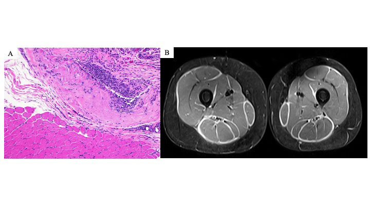
Methods: We report the demographics, clinical findings, pathology, MRIs, treatment, and outcomes for six patients with juvenile EF. A retrospective chart review was performed for all patients diagnosed with EF at our institution between November 2011- May 2022. Inclusion criteria included age at diagnosis < 18 years as well as EF confirmation by histology and MRI (Figure 1).

Results: For our juvenile EF cohort, the majority were female (83%) (Table 1). There were 50% Hispanic and 50% non-Hispanic patients. Age at diagnosis ranged from 4-16 years (median: 13) with associated follow-up ranging from 2 months to 12 years. Duration of symptoms prior to diagnosis ranged from 1 month to 1 year (median: 9 months). Associated medical conditions included localized scleroderma (50%), Hashimoto’s thyroiditis, 22q11 deletion, and acquired hemophilia A. Most patients presented with bilateral, progressive painful swelling and severe joint limitation mostly without any positive skin findings; one patient presented with unilateral involvement. All patients presented with a positive prayer sign (Figure 2). Initial treatment included corticosteroids and methotrexate. Other medications included hydroxychloroquine, immunoglobulin, mycophenolate mofetil, rituximab, and tocilizumab. Two patients had relapse of both morphea and fasciitis. Now, 83% are in remission with the remaining patient with resolved musculoskeletal symptoms and an improving factor 8 level after 2 months of treatment.

Conclusion: Juvenile EF can present with swelling and progressive induration without skin abnormalities. Unlike adult cohorts, there were no underlying malignancies or associations with significant trauma. Previous juvenile EF cohorts have described systemic involvement (hepatosplenomegaly, lymphadenopathy), which was not present within this cohort. Although non-specific, the prayer sign could be a helpful clinical finding to identify juvenile EF leading to early recognition and preventing long-term disabling outcomes.

Supporting image 1

Figure 1. Examples of eosinophilic fasciitis pathology and MRI findings. (A) Left lower leg muscle and fascia biopsy from patient 4 consistent with eosinophilic fasciitis. Hematoxylin and eosin (H &E) section (magnification, x100) showing fascia with underlying muscle. The fascia shows thickening and inflammatory cell infiltration (many macrophages and lymphocytes, few plasma cells, and rare eosinophils). (B) MRI bilateral thighs with and without contrast of patient 4 demonstrating extensive, symmetric fasciitis on STIR axial image.

Supporting image 2

Table 1. Juvenile eosinophilic fasciitis cohort patient characteristics. Bolded laboratory values are elevated above reference ranges.

Supporting image 3

Figure 2. Positive prayer sign at initial presentation in juvenile eosinophilic fasciitis. Here are examples of patients 3, 4, 5, and 6 demonstrating a positive prayer sign at the initial diagnosis. The prayer sign is due to skin induration and fascial fibrosis leading to joint contractures and tendon retraction.


Disclosures: L. Stubbs, None; O. Ogunbona, None; A. Adesina, None; S. Anvari, None; E. Beil, None; J. Lai, None; A. Ramirez, None; V. Szafron, None; M. Ditzler, None; M. DeGuzman, None.

To cite this abstract in AMA style:

Stubbs L, Ogunbona O, Adesina A, Anvari S, Beil E, Lai J, Ramirez A, Szafron V, Ditzler M, DeGuzman M. Juvenile Eosinophilic Fasciitis: A Single-Center Cohort [abstract]. Arthritis Rheumatol. 2022; 74 (suppl 9). https://acrabstracts.org/abstract/juvenile-eosinophilic-fasciitis-a-single-center-cohort/. Accessed .
  • Tweet
  • Email
  • Print

« Back to ACR Convergence 2022

ACR Meeting Abstracts - https://acrabstracts.org/abstract/juvenile-eosinophilic-fasciitis-a-single-center-cohort/

Advanced Search

Your Favorites

You can save and print a list of your favorite abstracts during your browser session by clicking the “Favorite” button at the bottom of any abstract. View your favorites »

All abstracts accepted to ACR Convergence are under media embargo once the ACR has notified presenters of their abstract’s acceptance. They may be presented at other meetings or published as manuscripts after this time but should not be discussed in non-scholarly venues or outlets. The following embargo policies are strictly enforced by the ACR.

Accepted abstracts are made available to the public online in advance of the meeting and are published in a special online supplement of our scientific journal, Arthritis & Rheumatology. Information contained in those abstracts may not be released until the abstracts appear online. In an exception to the media embargo, academic institutions, private organizations, and companies with products whose value may be influenced by information contained in an abstract may issue a press release to coincide with the availability of an ACR abstract on the ACR website. However, the ACR continues to require that information that goes beyond that contained in the abstract (e.g., discussion of the abstract done as part of editorial news coverage) is under media embargo until 10:00 AM ET on November 14, 2024. Journalists with access to embargoed information cannot release articles or editorial news coverage before this time. Editorial news coverage is considered original articles/videos developed by employed journalists to report facts, commentary, and subject matter expert quotes in a narrative form using a variety of sources (e.g., research, announcements, press releases, events, etc.).

Violation of this policy may result in the abstract being withdrawn from the meeting and other measures deemed appropriate. Authors are responsible for notifying colleagues, institutions, communications firms, and all other stakeholders related to the development or promotion of the abstract about this policy. If you have questions about the ACR abstract embargo policy, please contact ACR abstracts staff at [email protected].

Wiley

  • Online Journal
  • Privacy Policy
  • Permissions Policies
  • Cookie Preferences

© Copyright 2025 American College of Rheumatology