Session Information
Session Type: Poster Session B
Session Time: 8:30AM-10:30AM
Background/Purpose: Multiple diagnostic methods have been used to investigate muscle involvement in patients with idiopathic inflammatory myositis (IIM) such as electromyography (EMG) and muscle MRI. 18F-FDG PET-CT has been used to screen malignancy, and may also be beneficial in the investigation of myositis [1].
In this study, we aimed to evaluate the muscle involvement with PET – CT, which was performed for malignancy screening, and its association with clinical and laboratory parameters in patients with IIM.
Methods: IIM patients who fulfilled EULAR/ACR classification criteria and had PET-CT scans during the active phase of myositis included into the study. PET-CT scans of IIM patients and non-IIM patients with malignancy as controls were assessed retrospectively by two experienced nuclear medicine specialists and decisions were made by consensus. Positive PET-CT for myositis was defined as higher FDG muscle uptake compared to liver, mediastinal vascular structures (mediastinum) and lumbar longus muscle (LLM). Univariate and multivariate analysis were performed according to FDG uptake compared to mediastinum which was found to have higher diagnostic accuracy.
Results: One hundred and sixty IIM patients were screened and 34 patients (of 64.7 % female) whose PET-CT results were available and 14 non-IIM patients included into the study. Mean age of the IIM patients was 55±13 (25-85). Median disease duration of IIM patients was 12 months. Liver, mediastinum and LLM uptakes were similar in patients with IIM and non-IIM groups; higher muscle uptake was observed in proximal and distal upper (p=0.001, p< 0.001, respectively) and lower extremity (p=0.003, p< 0.001, respectively) in patients with IIM compared to non-IIM. Sensitivity and specificity of positive FDG muscle uptake were 37.1 % and 100 %, 65.7 % and 92.9 %, 91.4 % and 7.1 % compared to liver, mediastinum and LLM uptakes, respectively (Table 1).
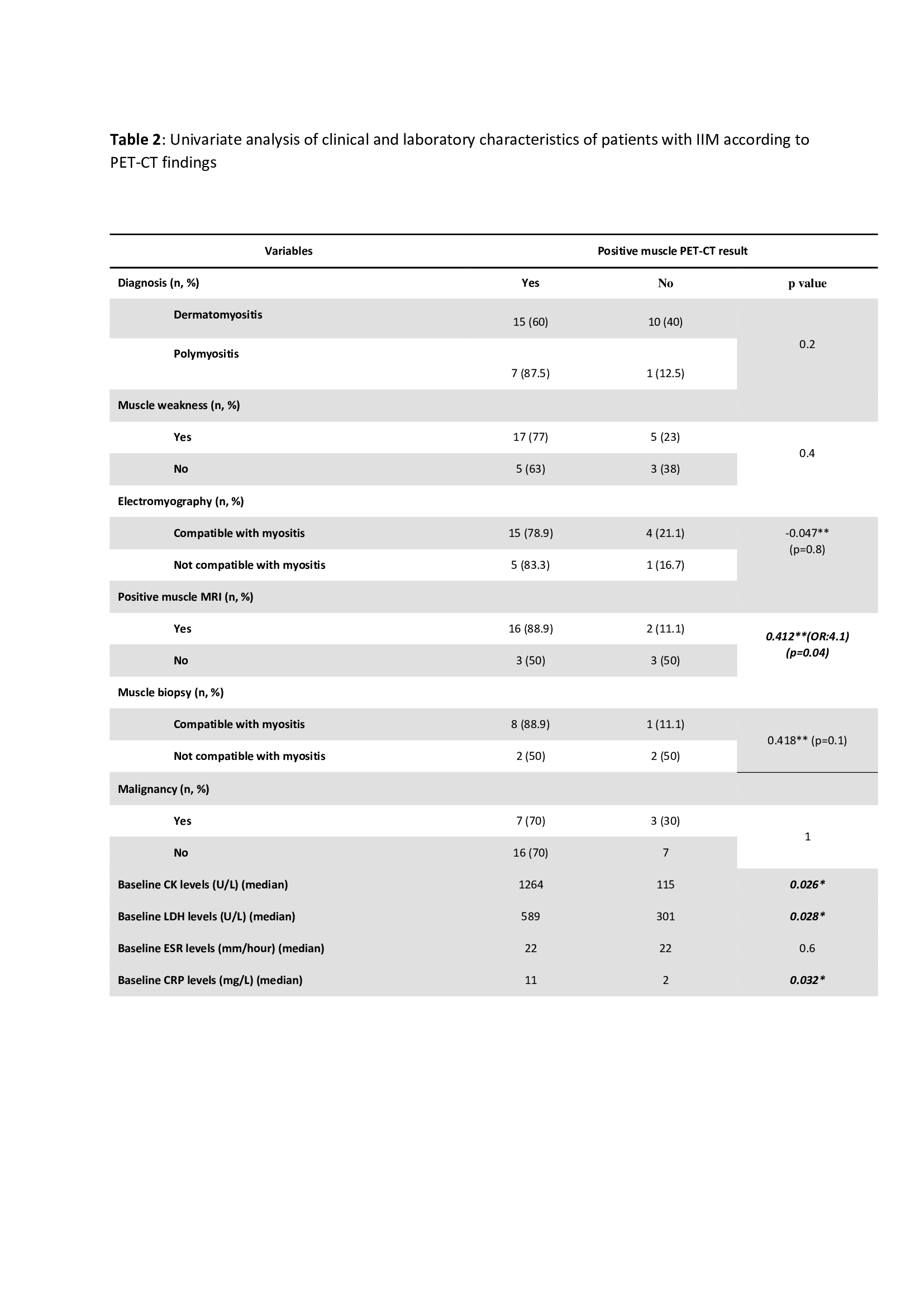
In univariate analysis; increased CK, LDH and CRP levels (p=0.026, p=0.028 and p=0.032 respectively) and positive muscle MRI (p=0.04; OR:4.1) were associated with PET-CT positivity. There was significant agreement between PET-CT positivity and muscle MRI findings (ĸ=0.412; p=0.04) (Table 2). In multivariate analysis, high baseline CRP (p=0.017, CI 95%: 1.03-1.36, OR:1.18) and LDH (p=0.029, CI 95%: 1.001-1.017, OR:1.01) levels were associated with PET-CT positivity. Baseline muscle strength (r=-0.411, p=0.04) was negatively correlated with muscle FDG uptake.
Conclusion: In patients with active IIM, median muscle FDG uptake with PET-CT was higher compared to non-IIM and significantly increased compared to liver and mediastinum. Muscle FDG values compared to mediastinum had the highest sensitivity. PET-CT findings are correlated with biomarkers of inflammation and clinical activity. PET-CT may be used for the evaluation of extent and activity in patients with IIM although further prospective research is needed.
1. Tateyama, M., et al., BMJ Open, 2015. 5(1): p. e006763

Ɨ Fisher’s exact test
IIM: Idiopathic inflammatory myositis, SD: Standard deviation, SUV-max: Standardized uptake value-maximum, PET-CT: Positron emission tomography, LLM: Lumbar longus muscle
SD: Standard deviation, CK: Creatinine kinase, LDH: Lactate dehydrogenase, ESR: Erythrocyte sedimentation rate, CRP: C-reactive protein, MRI: Magnetic resonance imaging
To cite this abstract in AMA style:
Bektaş M, Işık E, Kemik F, Oğuz E, ABBASGHOLI ZADEH A, Ince B, Gözde Özkan Z, Yalcinkaya Y, Esen B, Gül A, İnanç M. Is It Useful to Assess Muscle Involvement with Positron Emission Tomography in Patients with Idiopathic Inflammatory Myositis? A Case-Control Study [abstract]. Arthritis Rheumatol. 2021; 73 (suppl 9). https://acrabstracts.org/abstract/is-it-useful-to-assess-muscle-involvement-with-positron-emission-tomography-in-patients-with-idiopathic-inflammatory-myositis-a-case-control-study/. Accessed .« Back to ACR Convergence 2021
ACR Meeting Abstracts - https://acrabstracts.org/abstract/is-it-useful-to-assess-muscle-involvement-with-positron-emission-tomography-in-patients-with-idiopathic-inflammatory-myositis-a-case-control-study/