Session Information
Session Type: Poster Session B
Session Time: 10:30AM-12:30PM
Background/Purpose: There has been accumulating evidence that cellular senescence of fibroblasts and alveolar epithelial cells contributes to inflammation and fibrosis through a senescence-associated secretory phenotype in idiopathic pulmonary fibrosis. However, the associations between cellular senescence and connective tissue disease-associated interstitial lung diseases (ILD) including rheumatoid arthritis-associated ILD (RA-ILD), remains unknown. Therefore, we explored the involvement of cellular senescence in the pathogenesis of RA-ILD using SKG/Jcl mice.
Methods: We induced ILD in 10-week-old male SKG/Jcl mice (SKG-ILD) via intraperitoneal administration of zymosan, and dissected lungs after 4, 8, or 12 weeks. Senescent cells were detected by immunohistochemistry using cellular senescence markers, p21WAF1/CIP1 (p21), p16INK4a (p16), and γ-H2A histone family member X (γ-H2AX). To explore cell populations expressing p21, we performed immunofluorescence and flow cytometry using the antibodies against p21 in combination with cell surface markers for immune cells (CD45), endothelial cells (CD31), and epithelial cells (epithelial cell adhesion molecule; EpCAM). Single cell RNA sequencing (scRNA-seq) was performed on whole cell suspensions from SKG-ILD and control lungs.
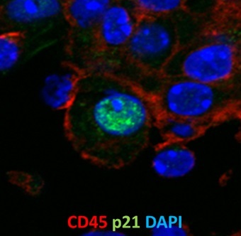
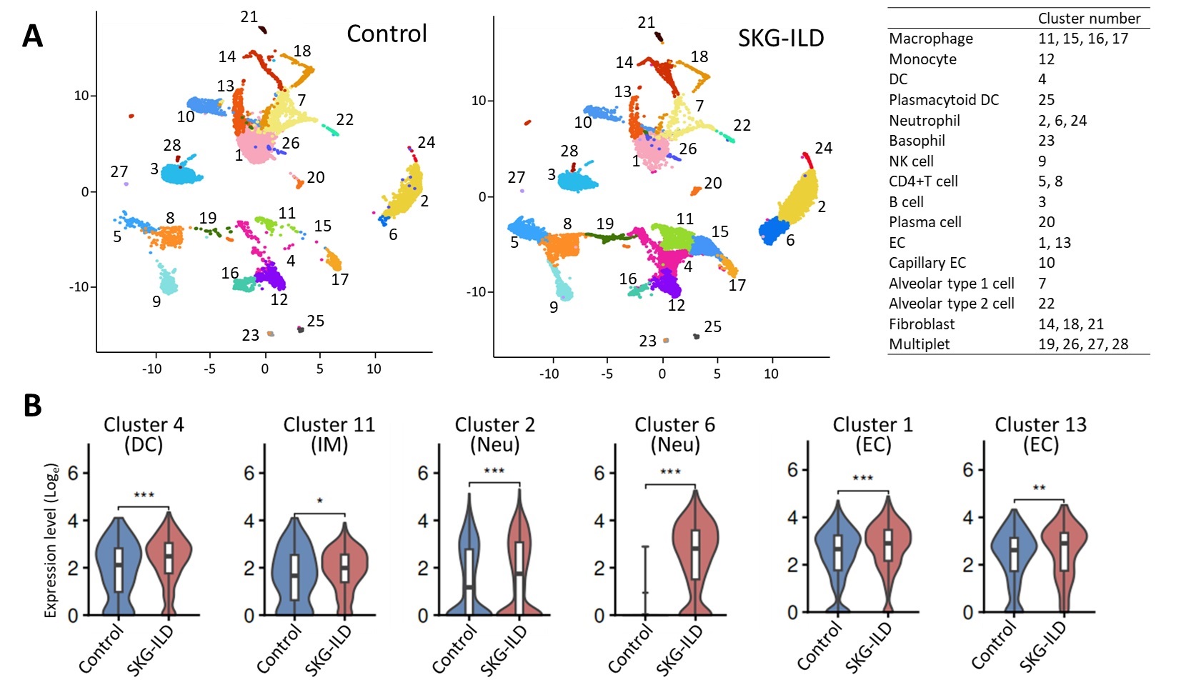
Results: In immunohistochemistry, p21-expressing cells appeared in SKG-ILD lungs at 4 weeks after administering zymosan, and increased with the progression of ILD. Twelve weeks post-administration, p21, p16 and γ-H2AX were localized in the fibrosis foci of SKG-ILD lungs, whereas the proliferation marker Ki-67 was negative. Immunofluorescence revealed that most p21-postive cells in the fibrotic foci were CD45+ cells (Figure 1), and some were CD31+ cells, whereas very few were EpCAM+ cells. Flow cytometry demonstrated that among cells expressing p21 in SKG-ILD lungs, the CD45+ cells showed the highest percentage. The scRNA-seq revealed significantly higher expression of Cdkn1a (the gene encoding p21) in myeloid cells including interstitial macrophages (cluster 11), dendritic cells (cluster 4) and neutrophils (clusters 2 and 6), and endothelial cells (clusters 1 and 13) in SKG-ILD lungs (Figure 2). Notably, cells in cluster 6, which were almost exclusively observed in SKG-ILD lungs and scarcely in control lungs, highly expressed interferon-stimulated genes such as Ifit3 and Isg15.
Conclusion: This study is the first to investigate the association of cellular senescence with RA-ILD using SKG-ILD. Our findings suggested that in SKG-ILD lungs, myeloid cells including interstitial macrophages, dendritic cells, neutrophils, and endothelial cells, may undergo cellular senescence, and play a role in the pathogenesis of RA-ILD.
SKG-ILD lung tissue was stained for CD45 (red), p21 (green), and DAPI (blue) by immunofluorescence.
(A) UMAP visualization for control and SKG-ILD lungs. (B) Expression of Cdkn1a in clusters 4, 11, 2, 6, 1 and 13.
DC, dendritic cell; IM, interstitial macrophage; Neu, neutrophil; EC, endothelial cell.
To cite this abstract in AMA style:
Watanabe E, Nishio J, Ban T, Yamada Z, Tamura T, Nanki T. Involvement of Cellular Senescence in Rheumatoid Arthritis-associated Interstitial Lung Disease of SKG Mice [abstract]. Arthritis Rheumatol. 2024; 76 (suppl 9). https://acrabstracts.org/abstract/involvement-of-cellular-senescence-in-rheumatoid-arthritis-associated-interstitial-lung-disease-of-skg-mice/. Accessed .« Back to ACR Convergence 2024
ACR Meeting Abstracts - https://acrabstracts.org/abstract/involvement-of-cellular-senescence-in-rheumatoid-arthritis-associated-interstitial-lung-disease-of-skg-mice/


