Session Information
Date: Monday, October 27, 2025
Title: (1147–1190) Miscellaneous Rheumatic & Inflammatory Diseases Poster II
Session Type: Poster Session B
Session Time: 10:30AM-12:30PM
Background/Purpose: Polymyalgia rheumatica (PMR) is characterized by increases in serum IL-6 and the proportion of Th17 cells in circulating blood. During PMR, IL-6 may originate from circulating monocytes or from the synovial tissue itself. To clarify the role of synovial tissue, we performed experiments using synovial fibroblast (SF) cultures derived from synovial bursae, which are the affected structures in PMR. As secukinumab (SEC) is currently being evaluated in glucocorticoid-dependent PMR, we investigated, in the current study, the impact of IL-17A and SEC on SFs activation.
Methods: SFs were obtained from ex-vivo cultures of synovial tissue from the subacromial bursae after arthroscopy in patients with rotator cuff injury. SFs were cultivated in vitro with IL-17A (50 ng/mL), IFN-g (2.5 ng/mL) and/or SEC (20 µg/mL) for 24 hours. mRNA expression was analyzed by Bulk RNA sequencing and RT-PCR. The impact of IL-17A on T-cell polarization was assessed by performing co-cultures of SFs and allogenic peripheral blood mononuclear cells (PBMC) activated with anti CD3/CD28 beads. SFs were stimulated with IL-17A or IL-17A + SEC or IgG isotype control for 72 hours, then co-cultures were performed over 7 days. T-cell polarization was analyzed by flow cytometry.
Results: The cells obtained through synovial tissue culture exhibited a phenotype fitting with fibroblasts (CD90+CD105+CD73intCD34-CD45-). Bulk RNA sequencing analysis showed increased levels of expression for genes associated with IL-17, PI3k-Akt and the NF-KB signaling pathway. Stimulation with IL-17A led to significant increases in 28 genes, notably IL6, CSF2 (encoding for GM-CSF), and CCL2. The addition of SEC led to a significant decrease in these mRNAs, restoring a transcriptomic profile similar to unstimulated SF (figure 1). RT-PCR analyses confirmed an increase in the mRNA expression of IL6, CSF2 and CCL2 in the presence of IL-17A, while no difference was observed for IL6ST, IFNGR1/2, IL12A, IL23A, FAP, IL17RA and IL17RC mRNA. Co-stimulation with IL-17A and IFN-g led to a strong increase in mRNA expression of IL6, IL6ST, and CCL2 (figure 2A). This synergy between IL-17A and IFN-g may be reliant on an increase in IL17RA mRNA expression after IFN-g stimulation (figure 2B). Co-cultures of SFs with allogenic PBMC were performed after 72h of SFs stimulation with either IgG isotype control, IL-17A or IL-17A+SEC. Compared to SFs pre-treated with IgG or IL-17A+SEC, SFs pre-treated with IL-17A were able to promote Th17 cell polarization without significant increases in the proportions of Th1, Tc1 or Tc17 cells (figure 3).
Conclusion: This work highlights the potential role of SFs in PMR via their ability to strongly express IL6. SFs are also sensitive to IL-17A and IFN-y, which are two key cytokines in PMR. In response to IL-17A, and with a synergistic effect with IFN-y, SFs strongly express IL6, leading to an increase in Th17 polarization. In addition, SFs express other markers such as CCL2, suggesting that they may induce the recruitment of CCR2+ monocytes. Finally, SEC can restore a non-inflammatory profile in activated SF, suggesting the potential of IL-17A therapeutic target in PMR.
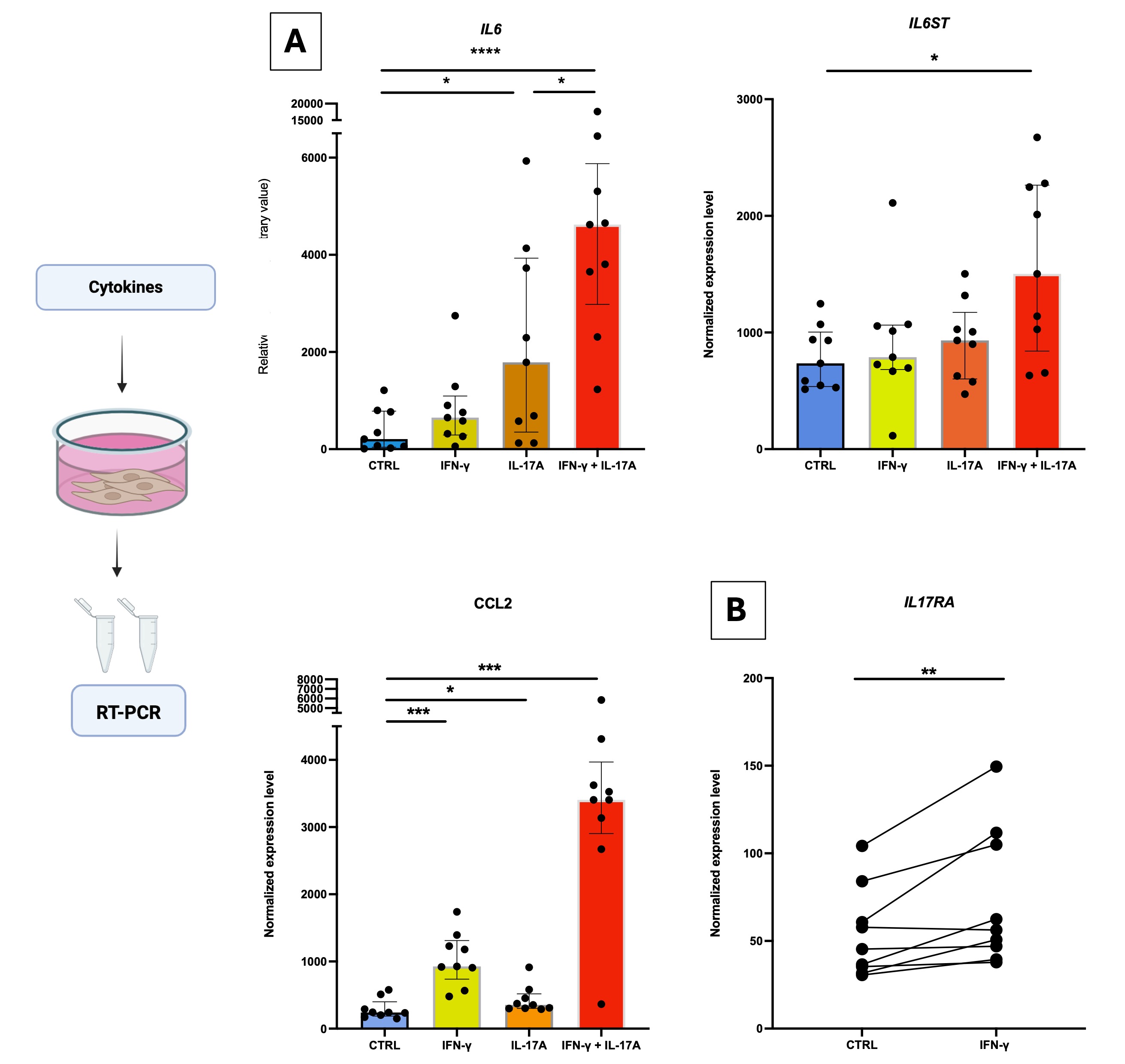 Figure 1: Bulk RNA sequencing results from IL-17A stimulated synovial fibroblasts (n = 8).
(A) Volcano plot showing differential gene expression (IL-17A versus IgG; IL-17A + SEC versus IgG).
(B) Barplot visualization of IL6, CSF2 and CCL2 gene expression from IL-17A, IL-17A + SEC and IgG stimulated synovial fibroblasts. Columns represent the median, the “whiskers” represent quartiles Q1 and Q3. *: p < 0.05, **: p < 0.01.
.jpg) Figure 2: Level of mRNA expression of IL6, IL6ST, CCL2 and IL17RA by synovial fibroblasts after stimulation with IL-17A and/or IFN-γ (n = 9).
Figure 2: Level of mRNA expression of IL6, IL6ST, CCL2 and IL17RA by synovial fibroblasts after stimulation with IL-17A and/or IFN-γ (n = 9).
A: Level of mRNA expression of IL6, IL6ST and CCL2 in synovial fibroblasts after stimulation with IL-17A (50 ng/mL) and/or IFN-γ (2,5 ng/mL). B: Effect of IFN-γ on mRNA expression of IL17RA by synovial fibroblasts. Columns represent the median, the “whiskers” represent quartiles Q1 and Q3. *: p < 0.05, **: p < 0.01, ***: p < 0.001, ****: p < 0.0001
.jpg) Figure 3: Study of the effect of synovial fibroblasts on T-cell polarization.
Figure 3: Study of the effect of synovial fibroblasts on T-cell polarization.
Percentage of Th1 (CD4+CD8-IFN-γ+), Tc1 (CD4-CD8+ IFN-γ-), Th17 (CD4+CD8-IL-17+), and Tc17 (CD4-CD8+IL-17+) cells. Columns represent the median, the “whiskers” represent quartiles Q1 and Q3. *: p < 0.05, ***: p < 0.001. NS = not significant. SEC: secukinumab. Coculture was performed using 8 samples of synovial tissue.
To cite this abstract in AMA style:
Ramon A, Greigert H, lamarthée b, Richard C, Cladière c, Klopfenstein N, Labattut L, Bordet A, Audia S, boidot R, Maillefert j, Bonnotte B, Samson M. Impact of IL-17A on synovial fibroblast from subacromial bursae [abstract]. Arthritis Rheumatol. 2025; 77 (suppl 9). https://acrabstracts.org/abstract/impact-of-il-17a-on-synovial-fibroblast-from-subacromial-bursae/. Accessed .« Back to ACR Convergence 2025
ACR Meeting Abstracts - https://acrabstracts.org/abstract/impact-of-il-17a-on-synovial-fibroblast-from-subacromial-bursae/