Session Information
Date: Monday, October 27, 2025
Session Type: Abstract Session
Session Time: 11:15AM-11:30AM
Background/Purpose: Most patients with inflammatory myopathies have autoantibodies to intracellular proteins. We previously showed that immunoglobulin colocalizes with its cognate autoantigen in muscle biopsies from myositis patients. Transcriptomic analyses of muscle biopsy specimens revealed that each myositis autoantibody is associated with a unique transcriptomic profile.We confirmed that these effects are antibody-mediated by using electroporation to internalize antibodies from patients into cultured muscle cells. Cultured muscle cells with internalized immunoglobulin from anti-MDA5+ and anti-Mi2+ patients expressed the same genes (e.g., Mx1 and CAMKV, respectively) as seen in the muscle tissue. The aims of the current study were to: (i) determine if electroporation of myositis-associated antibodies into human endothelial cells has similar transcriptomic profile as seen in muscle and (ii) to determine if immunoglobulin could passively enter cultured endothelial cells without electroporation.
Methods: Electroporation of purified antibody from Healthy controls, MDA5+, and Mi2 + sera was performed in HUVECS. The cells were incubated for 24hrs at 37 degrees Celsius following electroporation in appropriate growth media. The RNA was extracted from the HUVECs and RNA to CT was performed for comparative, quantitative PCR using Mx1 and CAMKV as probes and an18S as an internal loading control to assess gene expression.For passive antibody uptake experiments, HUVECs were cultured for 24 hours in growth media with purified healthy control, Mi2 +, and MDA5 + immunoglobin at 6 different concentrations (1:3 titration). RNA was the extracted using RNEAsy kits. Taqman RNA to CT kit was used to perform comparative, quantitative PCR to measure MX1 gene expression relative to an 18S loading control.
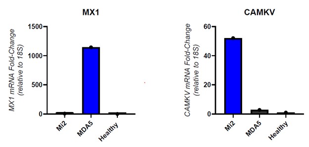
Results: Electroporation of MDA5 antibody increased resulted in increased relative MX1 expression as measured by PCR and electroporation of Mi2 antibodies resulted in relatively increased CAMKV compared to MDA5 and Healthy controls, respectively (Figure 1).Purified concentrated, MDA5 IgG that was passively introduced via a 24-hour incubation resulted in significantly increased relative Mx1 expression compared to healthy control IgG or Mi2 IgG (Figure 2).
Conclusion: These results suggest that autoantibodies are able to enter the cultured endothelial cells both with electroporation and passively, without electroporation. The higher relative MX1 expression with internalization of purified immunoglobulin from MDA5 and Mi2 compared to healthy controls suggests that the antibodies themselves may be contributing to pathogenesis and that endothelial cells may be contributing to disease pathogenesis in some types of idiopathic inflammatory myositis. Additional work is ongoing to elucidate the mechanism for the antibodies passively entering the cells and reaching the antigenic target.
.jpg)
To cite this abstract in AMA style:
Kinder T, Casal-Dominguez m, Pinal Fernandez I, Mammen A, Ogbonnaya-Whittlesey S. Immunoglobulin from sera of patients with myositis can passively enter cultured human endothelial cells [abstract]. Arthritis Rheumatol. 2025; 77 (suppl 9). https://acrabstracts.org/abstract/immunoglobulin-from-sera-of-patients-with-myositis-can-passively-enter-cultured-human-endothelial-cells/. Accessed .« Back to ACR Convergence 2025
ACR Meeting Abstracts - https://acrabstracts.org/abstract/immunoglobulin-from-sera-of-patients-with-myositis-can-passively-enter-cultured-human-endothelial-cells/