ACR Meeting Abstracts

ACR Meeting Abstracts

  • Meetings
    • ACR Convergence 2024
    • ACR Convergence 2023
    • 2023 ACR/ARP PRSYM
    • ACR Convergence 2022
    • ACR Convergence 2021
    • ACR Convergence 2020
    • 2020 ACR/ARP PRSYM
    • 2019 ACR/ARP Annual Meeting
    • 2018-2009 Meetings
    • Download Abstracts
  • Keyword Index
  • Advanced Search
  • Your Favorites
    • Favorites
    • Login
    • View and print all favorites
    • Clear all your favorites
  • ACR Meetings

Abstract Number: 1279

Imaging of Ankylosing Spondylitis By [18f]Fluoride PET-CT to Assess Bone Formation Activity and to Monitor Anti-TNF Therapy

Stefan Bruijnen1, Nicki Verweij2, Leonie van Duivenvoorde3, Dominique Baeten4, Christiaan van Denderen5, Joost Bot6, Otto Hoekstra7, Pieter Raijmakers6, Irene van der Horst - Bruinsma8 and Conny van der Laken2, 1Dept. of Rheumtaology, Amsterdam Rheumatology and immunology Center - location VU University Medical Center, Amsterdam, The Netherlands, Amsterdam, Netherlands, 2Dept. of Rheumatology, Amsterdam Rheumatology and immunology Center - location VU University Medical Center, Amsterdam, The Netherlands, Amsterdam, Netherlands, 3Amsterdam Rheumatology and immunology Center, Academic Medical Center, Amsterdam, Netherlands, 4Amsterdam Rheumatology and immunology Center, Amsterdam, Netherlands, 5READE, Amsterdam Rheumatology and immunology Center, Reade, Amsterdam, Netherlands, 6Department of Radiology & Nuclear Medicine, VU University Medical Center, Amsterdam, Netherlands, 7Nuclear medicine and PET research, VU University Medical Center, Amsterdam, Netherlands, 8Rheumatology, Amsterdam Rheumatology and immunology Center, VU University medical center, Amsterdam, Netherlands

Meeting: 2016 ACR/ARHP Annual Meeting

Date of first publication: September 28, 2016

Keywords: Ankylosing spondylitis (AS), anti-TNF therapy and positron emission tomography (PET), Imaging

  • Tweet
  • Click to email a link to a friend (Opens in new window) Email
  • Click to print (Opens in new window) Print
Session Information

Date: Monday, November 14, 2016

Title: Imaging of Rheumatic Diseases - Poster II: XR/CT/PET/MRI

Session Type: ACR Poster Session B

Session Time: 9:00AM-11:00AM

Background/Purpose: Bone formation is an important hallmark of ankylosing spondylitis (AS). Recently, we demonstrated that axial bone formation activity in AS patients can be visualized in vivo by [18F]Fluoride PET-CT. [18F]Fluoride PET-CT may therefore allow monitoring of changes of bone formation in AS during treatment. Therefore we investigated 1. changes in bone formation activity in AS patients that start anti-TNF treatment using  [18F]Fluoride-PET-CT and 2. Histology of PET identified lesions in spine (small feasibility sub-study).

Methods: We included 12 AS patients (female 7/12; age 39±11) with high disease activity (BASDAI 5.5±1.1). [18F]fluoride PET-CT scans were performed at baseline and 12 weeks of anti-TNF treatment. Two patients only obtained a baseline scan to identify PET+ lesions in spine for a subsequent  biopsy procedure to collect material of these lesions for immunohistochemistry (IHC). PET scans were assessed visually by two blinded readers. Clinical response was defined according to the ASAS20 at 12 weeks.

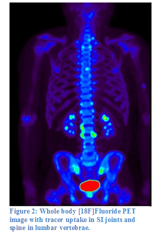
Results: Visualization of bone formation by [18F]fluoride PET-CT in spine was supported by osteoid formation and osteoclasts along with cell infiltrate in conjunction areas of bone and connective tissue in PET+ lesions, while largely absent in PET- lesions (Figure 1A,B). At baseline, the other 10 patients showed at least one lesion of [18F]Fluoride uptake in spine and/or SI joints. In spine, a total of 94 PET+ lesions were observed in 6 patients (range 2-32 p.p.), and ≥1 SI PET+ joint in 8/10 patients (Fig. 2). After 12 weeks of anti-TNF treatment, 7/10 patients received ASAS20 response. In spine, in 6/10pts with ≥1 PET+ lesion at baseline, the number of PET+ lesions decreased from 94 to 66 in 12 weeks. Decrease of spine+ lesions was noticed both in clinical responders and non-responders although the remaining number of spine+ lesions at 12 weeks tended to be higher in non-responders than in responders (16-21 vs. 1-13, respectively). SI+ joints remained visually positive at 12 weeks although at variable intensity.

Conclusion: In AS patients, [18F]Fluoride PET-CT visualizes multiple lesions with bone formation in SI joints and spine. Histological evaluation of PET+ lesions supported in-vivo identification of bone formation activity by [18F]Fluoride PET-CT. In this preliminary visual analysis of PET data, one third of [18F]Fluoride PET-CT+ lesions in spine disappeared after 12 weeks of a-TNF treatment. The relationship between PET outcome and clinical response will be further investigated by quantitative analysis of PET+ lesions and clinical follow-up up to 24 weeks, which is currently in progress. 


Disclosure: S. Bruijnen, Dutch Arthritis Association, 2; N. Verweij, None; L. van Duivenvoorde, None; D. Baeten, None; C. van Denderen, None; J. Bot, None; O. Hoekstra, None; P. Raijmakers, None; I. van der Horst - Bruinsma, None; C. van der Laken, None.

To cite this abstract in AMA style:

Bruijnen S, Verweij N, van Duivenvoorde L, Baeten D, van Denderen C, Bot J, Hoekstra O, Raijmakers P, van der Horst - Bruinsma I, van der Laken C. Imaging of Ankylosing Spondylitis By [18f]Fluoride PET-CT to Assess Bone Formation Activity and to Monitor Anti-TNF Therapy [abstract]. Arthritis Rheumatol. 2016; 68 (suppl 10). https://acrabstracts.org/abstract/imaging-of-ankylosing-spondylitis-by-18ffluoride-pet-ct-to-assess-bone-formation-activity-and-to-monitor-anti-tnf-therapy/. Accessed .
  • Tweet
  • Click to email a link to a friend (Opens in new window) Email
  • Click to print (Opens in new window) Print

« Back to 2016 ACR/ARHP Annual Meeting

ACR Meeting Abstracts - https://acrabstracts.org/abstract/imaging-of-ankylosing-spondylitis-by-18ffluoride-pet-ct-to-assess-bone-formation-activity-and-to-monitor-anti-tnf-therapy/

Advanced Search

Your Favorites

You can save and print a list of your favorite abstracts during your browser session by clicking the “Favorite” button at the bottom of any abstract. View your favorites »

All abstracts accepted to ACR Convergence are under media embargo once the ACR has notified presenters of their abstract’s acceptance. They may be presented at other meetings or published as manuscripts after this time but should not be discussed in non-scholarly venues or outlets. The following embargo policies are strictly enforced by the ACR.

Accepted abstracts are made available to the public online in advance of the meeting and are published in a special online supplement of our scientific journal, Arthritis & Rheumatology. Information contained in those abstracts may not be released until the abstracts appear online. In an exception to the media embargo, academic institutions, private organizations, and companies with products whose value may be influenced by information contained in an abstract may issue a press release to coincide with the availability of an ACR abstract on the ACR website. However, the ACR continues to require that information that goes beyond that contained in the abstract (e.g., discussion of the abstract done as part of editorial news coverage) is under media embargo until 10:00 AM ET on November 14, 2024. Journalists with access to embargoed information cannot release articles or editorial news coverage before this time. Editorial news coverage is considered original articles/videos developed by employed journalists to report facts, commentary, and subject matter expert quotes in a narrative form using a variety of sources (e.g., research, announcements, press releases, events, etc.).

Violation of this policy may result in the abstract being withdrawn from the meeting and other measures deemed appropriate. Authors are responsible for notifying colleagues, institutions, communications firms, and all other stakeholders related to the development or promotion of the abstract about this policy. If you have questions about the ACR abstract embargo policy, please contact ACR abstracts staff at [email protected].

Wiley

  • Online Journal
  • Privacy Policy
  • Permissions Policies
  • Cookie Preferences

© Copyright 2025 American College of Rheumatology