Session Information
Session Type: Poster Session B
Session Time: 9:00AM-11:00AM
Background/Purpose: We previously discovered that IL-16 is the urinary protein most strongly correlated with histological activity in lupus nephritis (LN), followed by neutrophil degranulation products such as PR3. IL-16 is a proinflammatory chemokine that binds to CD4 and CD9 which are also expressedby myeloid cells. IL-16 is a key mediator in renal ischemia-reperfusion injury. Furthermore, IL-16 polymorphisms carry a higher risk of SLE (OR 3.3-10.4), suggesting a potential causal role. Our hypothesis is that IL-16 critically amplifies the immune response in proliferative LN, thereby attracting myeloid populations that can ultimately lead to kidney damage. The aims of our study are to investigate IL-16+ cells in LN kidney, define their immunophenotype, and to explore the presence of PR3+ cells in the same spatial area in order to gain insight on the spatial relation between the two populations.
Methods: We performed 20-plex serial immunohistochemistry (sIHC) on one archival LN kidney biopsy (ISN/RPS class IV) to identify IL-16+ and PR3+ cells and evaluate the expression of multiple cell lineage markers. We utilized Indica HALO to perform image analysis, including deconvolution, cell segmentation, glomerular annotation, and quantitative histology.
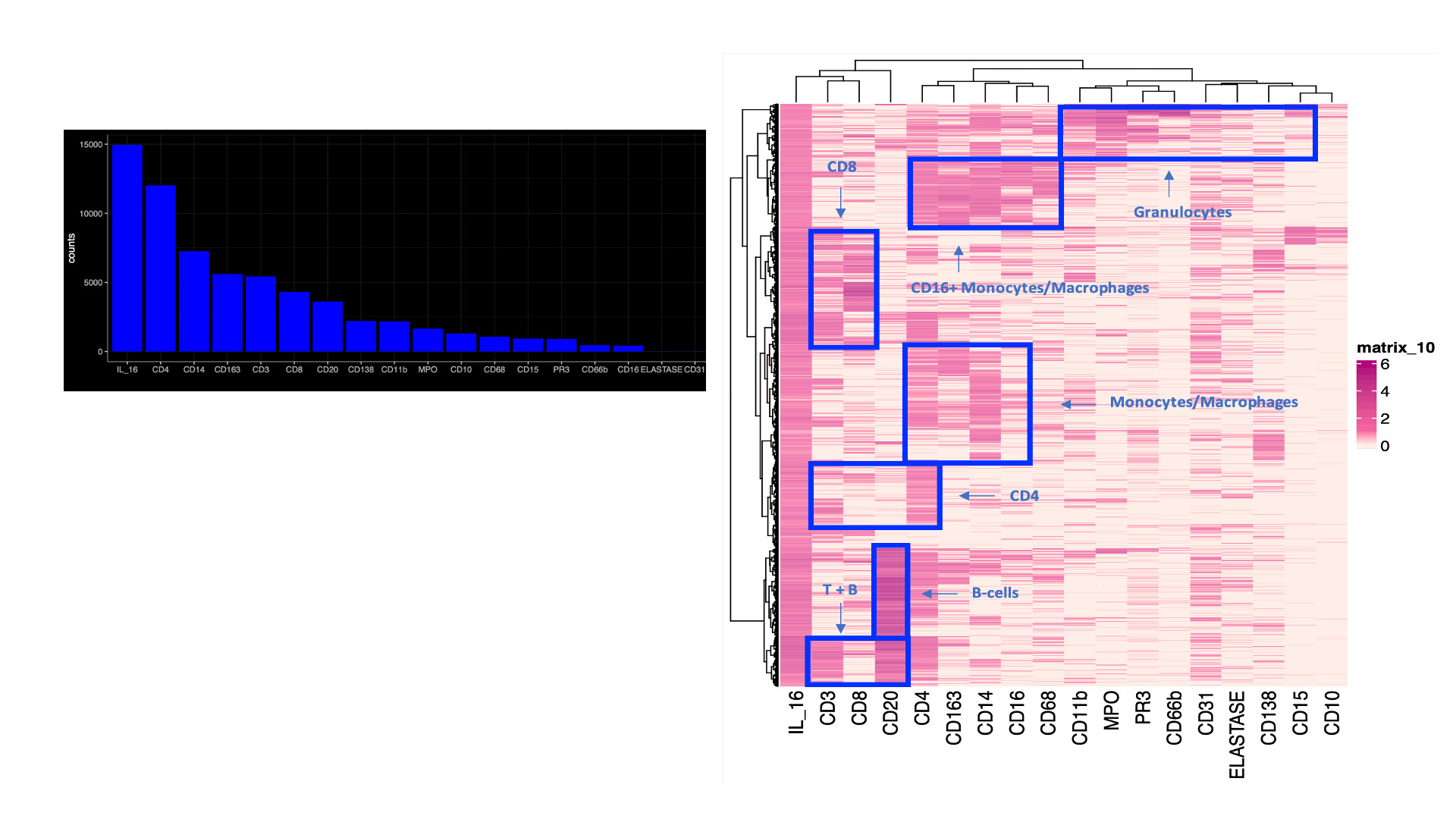
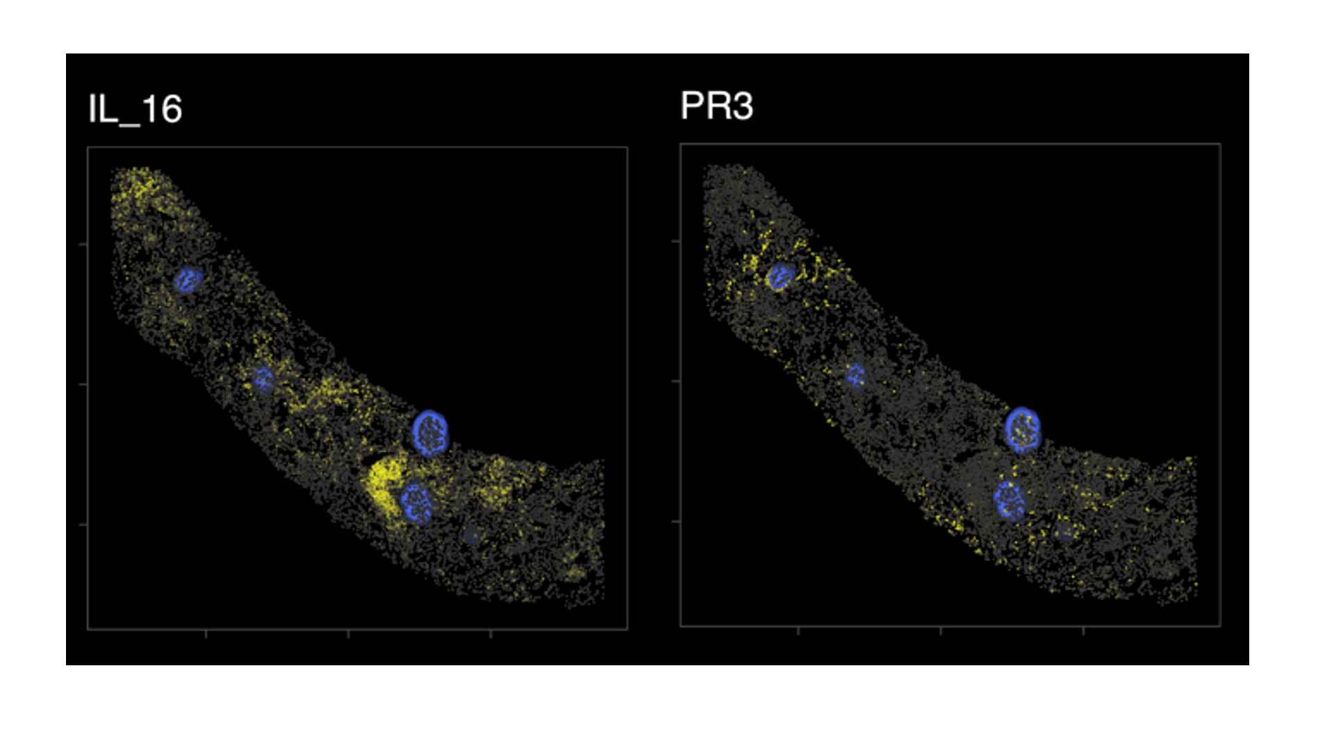
Results: A total of 95,619 cell objects present in the sample were analyzed. We observed an abundant IL-16+ infiltrate, mostly in the tubulointerstitium. Multiple kidney-infiltrating immune cell types produced IL-16. The co-expression of lineage markers by IL-16+ cells is summarized by Figure1A. Figure 1Bdisplays the heterogeneity of marker co-expression at the single cell level for IL-16+ cells. Most IL-16+ cells also expressed CD4, a ligand for IL-16. Figure 2 illustrates the tissue distribution of IL-16 and PR3+ cells showing how IL-16 and PR3 are not spatially related.
Conclusion: IL-16+ cells abundantly infiltrate the kidney in LN. IL-16 is producedby most types of immune cells including lymphoid and myeloid cells. There was no spatial relationship between IL-16+ and PR3+ cells suggesting no obvious chemotactic activity. The release of IL-16 by multiple immune cell types may promote and amplify LN.
(A) Coexpression of lineage markers in IL_16+ cells. Values indicate number (count) of positive cells. (B) Heatmaps displaying the heterogeneity of marker coexpression at the single cell level for IL_16+ cells (B). Data from one representative kidney biopsy (class IV lupus nephritis, NIH Activity Index 8/24, Chronicity Index 6/12).
Biopsy detail mapping IL_16 and PR3+ cells (in yellow); glomeruli in blue. There was no obvious spatial relation between IL16+ and PR3+ cells. Data from one representative kidney biopsy (class IV lupus nephritis, NIH Activity Index 8/24, Chronicity Index 6/12).
To cite this abstract in AMA style:
Celia A, Yang X, Minsky H, Malvica S, Petri M, Accelerating Medicines Partnership in RA/SLE t, Rosenberg A, Fava A. IL-16+ Is Abundantly Expressed by Kidney-infiltrating Myeloid and Lymphoid Cells in Lupus Nephritis: A Spatially Resolved Multiplexed Approach [abstract]. Arthritis Rheumatol. 2023; 75 (suppl 9). https://acrabstracts.org/abstract/il-16-is-abundantly-expressed-by-kidney-infiltrating-myeloid-and-lymphoid-cells-in-lupus-nephritis-a-spatially-resolved-multiplexed-approach/. Accessed .« Back to ACR Convergence 2023
ACR Meeting Abstracts - https://acrabstracts.org/abstract/il-16-is-abundantly-expressed-by-kidney-infiltrating-myeloid-and-lymphoid-cells-in-lupus-nephritis-a-spatially-resolved-multiplexed-approach/


