ACR Meeting Abstracts

ACR Meeting Abstracts

  • Home
  • Meetings Archive
    • ACR Convergence 2021
    • ACR Convergence 2020
    • 2020 ACR/ARP PRSYM
    • 2019 ACR/ARP Annual Meeting
    • 2018 ACR/ARHP Annual Meeting
    • 2017 ACR/ARHP Annual Meeting
    • 2017 ACR/ARHP PRSYM
    • 2016-2009 Meetings
    • Download Abstracts
  • Keyword Index
  • Advanced Search
  • Your Favorites
    • Favorites
    • Login
    • View and print all favorites
    • Clear all your favorites
  • Meeting Resource Center

Abstract Number: 964

Identifying Jo-1-Specific B Cells in the Primary Immune Repertoire in Idiopathic Inflammatory Myopathies

Jennifer Young-Glazer1, Erin Wilfong 2, Peggy Kendall 1, Rachel Bonami 1 and Leslie Crofford 2, 1Vanderbilt University Medical Center, Nashville, TN, 2Vanderbilt University, Nashville

Meeting: 2019 ACR/ARP Annual Meeting

Keywords: Antibodies, B cells, polymyositis/dermatomyositis (PM/DM) and Idiopathic Inflammatory Myopathies (IIM), synthetase syndrome

  • Tweet
  • Email
  • Print
Session Information

Date: Monday, November 11, 2019

Session Title: B Cell Biology & Targets In Autoimmune & Inflammatory Disease Poster

Session Type: Poster Session (Monday)

Session Time: 9:00AM-11:00AM

Background/Purpose: Idiopathic inflammatory myopathies (IIM) are systemic autoimmune diseases traditionally classified as dermatomyositis or polymyositis, but these disorders are increasingly defined by the presence of myositis-specific and myositis-associated antibodies. Anti-Jo-1 autoantibodies recognize histidyl tRNA synthetase and are frequently found in these patients. In addition to producing antibody, B cells present antigen to T cells, a function that is independent of antibody production. Jo-1-specific B cells have not been previously characterized. To develop targeted therapies for IIM, we must first understand the etiology of autoreactive B cells in this disease. We hypothesize that anti-Jo-1 B cells will exhibit features of memory B cells and show evidence of somatic hypermutation and antigen-driven selection.

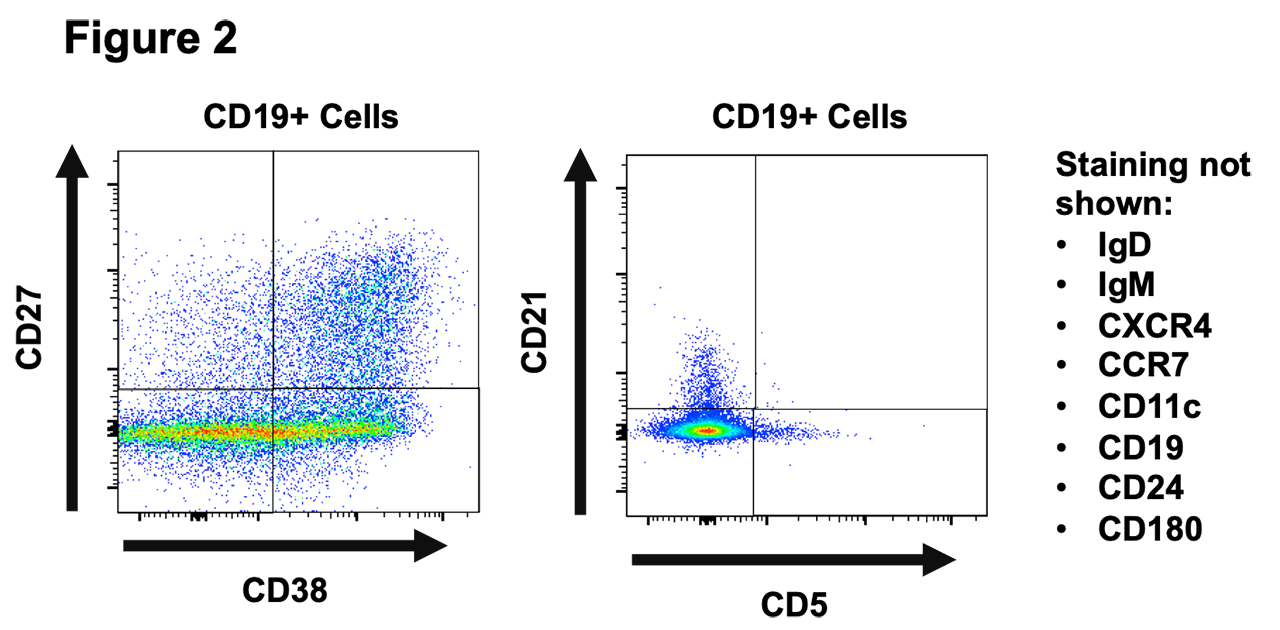
Methods: We enrolled 10 patients that were anti-Jo-1 autoantibody positive by clinical testing at our university medical center (Table 1). Antigen-specific B lymphocytes are typically rare in the repertoire and the B cell receptor is not automatically secreted as antibody, making detection of specificity difficult. To overcome these challenges, we polyclonally stimulated peripheral blood mononuclear cells (PBMCs) from IIM patients to drive in vitro B cell differentiation into antibody-secreting cells and developed a high throughput ELISA to screen and identify wells containing Jo-1 antibody-secreting cells. We applied flow cytometry phenotyping of wells that were positive or negative for anti-Jo-1 B to investigate their expression of twelve B cell phenotypic markers.

Results: Using recombinant Jo-1 protein, our ELISA successfully distinguished the presence or absence of anti-Jo-1 antibodies in clinically validated patient sera (Figure 1). Stimulation of human PBMC resulted in detection of IgG in the supernatants of all wells. A proportion of these wells were positive for anti-Jo-1 IgG autoantibody with an estimated frequency of approximately 0.01% of the total B cell repertoire, a frequency that is comparable to previously reported tetanus toxoid B cells in vaccinated individuals. We also detected a residual population of anti-Jo-1 B cells in a patient who previously received rituximab. B cell subsets differentially expressed CD24, CD27, CD38, IgM, and IgD following polyclonal stimulation. Differences in CXCR4 and CCR7 expression were also observed in different B cell subsets identified in these cultures.

Conclusion: This is the first demonstration of anti-Jo-1 B cells in the primary immune repertoire of patients with IIM, including those who have previously received rituximab. Flow phenotypic analysis suggests that differences in B cell subset distribution of anti-Jo-1 B cells can be assessed using our methods. These data provide the first critical steps towards understanding anti-Jo-1 B cell biology in patients with IIM to enable their future tracking as clinical disease and therapeutic response biomarkers.

Table 1: Demographics of patients and healthy volunteers included in study. Four healthy controls, 4 patients with non-Jo-1 inflammatory myopathy patients -one each with anti-Tif1-gamma, anti-Mi-2, anti-EJ, and anti-PL-7 antibodies-, and 10 patients with anti-Jo-1 antibodies as determined by clinical testing. Demographics and clinical information were collected from the electronic medical record at the time of enrollment. Disease severity was defined as stable if the patient’s primary rheumatologist or pulmonologist did not discuss changing medications at the time of enrollment and “active” if immunosuppression was increased. Disease activity was determined to be “life threatening” if the patient was admitted to the hospital due to their rheumatologic illness, was receiving pulse steroids, and was receiving an increase in immunosuppression.

Figure 1: ELISA detects anti-Jo-1 specific antibody in the sera of patients with clinical laboratory confirmed cases of Jo-1 associated disease. -A- Glutathione S-transferase -GST- and GST-Jo-1 were expressed in bacteria and purified. Samples were run on SDS-page gels and Coomassie blue staining confirmed purity of protein samples. -B- ELISA performed against purified GST-Jo-1 fusion protein -black- and purified GST protein -gray-. Patient sera were diluted 1:1000 and applied to plates coated with purified GST-Jo-1 or GST proteins. Our ELISA successfully discriminates between Jo-1-positive and Jo-1-negative sera. The Jo-1-positive patient sera react against GST-Jo-1 fusion protein but not against GST alone, demonstrating that the antibodies are specific for the Jo-1 protein.

Figure 2: Representative flow cytometry data. Cryopreserved PBMCs from a known Jo-1 positive patient were thawed and stimulated in vitro. After 6 days of stimulation, cell culture supernatants were screened for the presence of anti-Jo-1 antibodies. Equal numbers of Jo-1 positive and Jo-1 negative screened wells were phenotyped using flow cytometry. Representative data for a Jo-1 positive well are shown above amongst CD19+ cells.


Disclosure: J. Young-Glazer, None; E. Wilfong, None; P. Kendall, None; R. Bonami, None; L. Crofford, None.

To cite this abstract in AMA style:

Young-Glazer J, Wilfong E, Kendall P, Bonami R, Crofford L. Identifying Jo-1-Specific B Cells in the Primary Immune Repertoire in Idiopathic Inflammatory Myopathies [abstract]. Arthritis Rheumatol. 2019; 71 (suppl 10). https://acrabstracts.org/abstract/identifying-jo-1-specific-b-cells-in-the-primary-immune-repertoire-in-idiopathic-inflammatory-myopathies/. Accessed May 16, 2022.
  • Tweet
  • Email
  • Print

« Back to 2019 ACR/ARP Annual Meeting

ACR Meeting Abstracts - https://acrabstracts.org/abstract/identifying-jo-1-specific-b-cells-in-the-primary-immune-repertoire-in-idiopathic-inflammatory-myopathies/

Advanced Search

Your Favorites

You can save and print a list of your favorite abstracts during your browser session by clicking the “Favorite” button at the bottom of any abstract. View your favorites »

ACR Pediatric Rheumatology Symposium 2020

© COPYRIGHT 2022 AMERICAN COLLEGE OF RHEUMATOLOGY

Wiley

  • Home
  • Meetings Archive
  • Advanced Search
  • Meeting Resource Center
  • Online Journal
  • Privacy Policy
  • Permissions Policies
loading Cancel
Post was not sent - check your email addresses!
Email check failed, please try again
Sorry, your blog cannot share posts by email.