ACR Meeting Abstracts

ACR Meeting Abstracts

  • Meetings
    • ACR Convergence 2024
    • ACR Convergence 2023
    • 2023 ACR/ARP PRSYM
    • ACR Convergence 2022
    • ACR Convergence 2021
    • ACR Convergence 2020
    • 2020 ACR/ARP PRSYM
    • 2019 ACR/ARP Annual Meeting
    • 2018-2009 Meetings
    • Download Abstracts
  • Keyword Index
  • Advanced Search
  • Your Favorites
    • Favorites
    • Login
    • View and print all favorites
    • Clear all your favorites
  • ACR Meetings

Abstract Number: 0051

Identification of a Transcription Factor That Drives Polarization Toward Tissue-destructive Fibroblasts in Arthritis

MINGLU YAN1, Noriko Komatsu1, Ryunosuke Muro1, Hiroyuki Takaba1, Takeshi Nitta1, Kazuo Okamoto1, Masayuki Tsukasaki2 and Hiroshi Takayanagi1, 1University of Tokyo, Tokyo, Japan, 2The Univerisity of Tokyo, Tokyo, Japan

Meeting: ACR Convergence 2022

Keywords: Fibroblasts, Synovial, rheumatoid arthritis

  • Tweet
  • Click to email a link to a friend (Opens in new window) Email
  • Click to print (Opens in new window) Print
Session Information

Date: Saturday, November 12, 2022

Title: RA – Animal Models Poster

Session Type: Poster Session A

Session Time: 1:00PM-3:00PM

Background/Purpose: Fibroblasts exert important homeostatic functions but can also drive disease pathogenesis. In rheumatoid arthritis (RA), synovial fibroblasts (SFs) contribute to the joint destruction by producing the osteoclast differentiation factor receptor activator of nuclear factor-κB ligand RANKL as well as matrix metalloproteinases (MMPs) to induce bone erosion and cartilage damage, respectively. However, the mechanisms underlying the tissue-destructive fibroblast phenotype remain unclear.

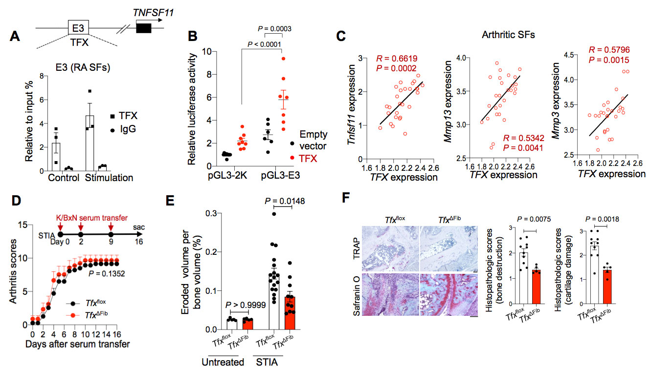
Methods: We performed single-cell RNA-sequencing (scRNA-seq) and epigenomic analyses of arthritic SFs to study the transcriptional mechanism underlying the tissue-destructive gene expression in SFs. Luciferase assay was performed to examine the functions of candidate enhancer elements and transcription factor (TFX) in vitro. We further generated the enhancer knockout (KO) mice and fibroblast-specificTFXconditional knockout mice and analyzed their phenotypes under steady state and arthritic conditions.

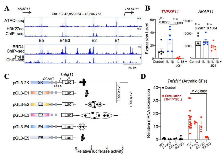
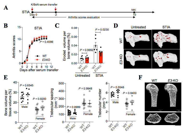
Results: Epigenomic analysis of arthritic SFs identified five distal enhancer elements (E1 to E5) upstream of the RANKL gene locus. Among the homologous regions of the five elements in mice, E3 was found to be capable of efficiently inducing RANKL transcriptional activity in an in vitro reporter assay. We further identified transcription factor TFX binds to E3 region and further enhanced RANKL gene transcription. Mice lacking E3 region displayed a reduced joint damage with a decreased RANKL expression in SFs under arthritic conditions, while retaining normal bone metabolism at steady state. Fibroblast-specific TFX deletion resulted in ameliorated bone and cartilage damage under arthritic conditions without affecting the inflammation.

Conclusion: Therefore, TFX drives the polarization toward tissue-destructive fibroblasts in arthritis. These findings provide a mechanistic basis for pathogenic fibroblast polarization in arthritis and have important clinical implications.

Supporting image 1

Fig. 1: A distal enhancer element regulates RANKL gene expression in arthritic SFs. A, Epigenomic analyses of SFs from RA patients at the TNFSF11 locus (hg19) by ATAC–seq (GSE128644), H3K27ac ChIP–seq (GSE128642), BRD4 ChIP–seq and Pol II ChIP–seq (GSE148399). B, The TNFSF11 (left) and AKAP11 (right) expression levels in RA patient-derived SFs treated with or without IL_1 and JQ1 (GSE148395). P values were determined by one-way ANOVA analysis followed by Turkey’s post hoc test. C, Reporter assay for the measurement of mouse E1-E5 enhancer activity for RANKL gene transcription. P values were determined by one-way ANOVA analysis followed by Turkey’s post hoc test. D, Tnfsf11 mRNA expression in the arthritic SFs stimulated with or without TNF/PGE2. P values were determined by two-way ANOVA analysis followed by Turkey’s post hoc test.

Supporting image 2

Fig.2: E3 deletion ameliorates arthritis-induced bone damage. A, Schematic showing murine serum transfer-induced arthritis (STIA) model. B, Arthritis scores of WT and E3-KO mice under STIA condition. P values were determined by two-tailed Mann-Whitney U-test. C, Eroded volume per bone volume of the ankle joints in WT and E3-KO mice under untreated and STIA (day 10) conditions analyzed by micro-CT. P values were determined by two-way ANOVA analysis followed by Turkey’s post hoc test. D, Representative micro-CT images of the ankles of WT and E3-KO mice under untreated and STIA conditions. The red area indicated the erosive cavities evaluated by micro-CT analysis. E, Micro-CT analysis of bone volume per tissue volume, trabecular spacing and number of WT and E3-KO mice at the age of 10 weeks (n=5_13) under physiological conditions. P values were determined by two-way ANOVA analysis followed by Turkey’s post hoc test. F, Representative micro-CT images of the femurs of WT and E3-KO male mice at the age of 10 weeks, micro-CT scale bars: 1 mm.

Supporting image 3

Fig. 3: TFX deletion in SFs attenuates arthritic joint damage. A, Schematic depicting the E3 region upstream of the TNFSF11 transcription start site and ChIP of TFX binding in the E3 region analyzed by q-PCR in RA patient SFs (with or without TNF/PGE2 stimulation). Data were expressed as mean ± s.e.m. B, Reporter assay for the measurement of the mouse E3 enhancer activity for RANKL gene transcription. P values were determined by two-way ANOVA analysis followed by Turkey’s post hoc test. C, Spearman’s correlation analysis between TFX expression and Tnfsf11, Mmp13 and Mmp3 expression in arthritic SFs isolated from STIA mice (GSE129451). D, Arthritis scores of TFX-flox mice and TFX-ΔFib mice under STIA condition. E, Eroded volume per bone volume of the ankle joints of TFX-flox and TFX-ΔFib mice under untreated and STIA conditions as analyzed by micro-CT. P values were determined by two-way ANOVA analysis followed by Turkey’s post hoc test. F, Representative TRAP and Safranin O staining images of the histological sections of ankle joints and the histopathological scores for bone destruction and cartilage damage under STIA condition. P values were determined by two-tailed t-test.


Disclosures: M. YAN, None; N. Komatsu, None; R. Muro, None; H. Takaba, None; T. Nitta, None; K. Okamoto, None; M. Tsukasaki, None; H. Takayanagi, None.

To cite this abstract in AMA style:

YAN M, Komatsu N, Muro R, Takaba H, Nitta T, Okamoto K, Tsukasaki M, Takayanagi H. Identification of a Transcription Factor That Drives Polarization Toward Tissue-destructive Fibroblasts in Arthritis [abstract]. Arthritis Rheumatol. 2022; 74 (suppl 9). https://acrabstracts.org/abstract/identification-of-a-transcription-factor-that-drives-polarization-toward-tissue-destructive-fibroblasts-in-arthritis/. Accessed .
  • Tweet
  • Click to email a link to a friend (Opens in new window) Email
  • Click to print (Opens in new window) Print

« Back to ACR Convergence 2022

ACR Meeting Abstracts - https://acrabstracts.org/abstract/identification-of-a-transcription-factor-that-drives-polarization-toward-tissue-destructive-fibroblasts-in-arthritis/

Advanced Search

Your Favorites

You can save and print a list of your favorite abstracts during your browser session by clicking the “Favorite” button at the bottom of any abstract. View your favorites »

All abstracts accepted to ACR Convergence are under media embargo once the ACR has notified presenters of their abstract’s acceptance. They may be presented at other meetings or published as manuscripts after this time but should not be discussed in non-scholarly venues or outlets. The following embargo policies are strictly enforced by the ACR.

Accepted abstracts are made available to the public online in advance of the meeting and are published in a special online supplement of our scientific journal, Arthritis & Rheumatology. Information contained in those abstracts may not be released until the abstracts appear online. In an exception to the media embargo, academic institutions, private organizations, and companies with products whose value may be influenced by information contained in an abstract may issue a press release to coincide with the availability of an ACR abstract on the ACR website. However, the ACR continues to require that information that goes beyond that contained in the abstract (e.g., discussion of the abstract done as part of editorial news coverage) is under media embargo until 10:00 AM ET on November 14, 2024. Journalists with access to embargoed information cannot release articles or editorial news coverage before this time. Editorial news coverage is considered original articles/videos developed by employed journalists to report facts, commentary, and subject matter expert quotes in a narrative form using a variety of sources (e.g., research, announcements, press releases, events, etc.).

Violation of this policy may result in the abstract being withdrawn from the meeting and other measures deemed appropriate. Authors are responsible for notifying colleagues, institutions, communications firms, and all other stakeholders related to the development or promotion of the abstract about this policy. If you have questions about the ACR abstract embargo policy, please contact ACR abstracts staff at [email protected].

Wiley

  • Online Journal
  • Privacy Policy
  • Permissions Policies
  • Cookie Preferences

© Copyright 2025 American College of Rheumatology