Session Information
Session Type: Poster Session C
Session Time: 10:30AM-12:30PM
Background/Purpose: Visual diagnosis, based on both physical exam and imaging studies, is of great importance in clinical practice and for board exam purposes in rheumatology. The rarity of many rheumatologic diseases limits fellow exposure to the full range of physical and radiologic disease manifestations during training. There is a continued need to develop high-quality learning materials within rheumatology training programs. We report on a quality improvement education project which aims to develop and continually improve a question-based visual diagnosis curriculum.
Methods: Visual diagnosis questions were created using the American College of Rheumatology Image Library as well as clinical cases at a single academic center. Topics were chosen based on relevance with a focus on rheumatoid arthritis, dermatomyositis, systemic lupus erythematosus, crystalline arthropathy and spondylarthritis. Questions were created by two fellows and reviewed by faculty for content accuracy. Visual diagnostic modalities included physical exam findings, plain radiographs, and advanced imaging such as MRI and CT. Five separate modules consisting of seven questions each were delivered to six fellows between March 21, 2025 and May 5, 2025 via Qualtrics. Survey questions were embedded into each module and feedback incorporated into the next PDSA cycle.
Results: Five to six fellows completed each module. Project aims, interventions, and evaluation are outlined in Figure 1. Continuous improvement of the modules based on focused areas of feedback took place throughout the project period as noted in Figure 2. Fellows demonstrated a measurable increase in confidence from pre-module to post-module assessments (Figure 3). Prior to module dissemination, fellows rated level of satisfaction with visual diagnosis curriculum as “somewhat satisfied” (n=3) or “neutral” (n=2). Conversely, all respondents (n=5) were “very satisfied” with the module questions for their training and preferred to continue this as a part of the fellowship curriculum.
Conclusion: There were high levels of engagement in this visual diagnosis curriculum utilizing fellow-generated, faculty-reviewed questions. Integration of feedback with curricular material for rapid changes into the curriculum may serve as a model for future educational quality improvement. Next steps include expanding topics to include additional imaging modalities such as ultrasound and diagnoses such as vasculitis, increasing the number of questions for expansion as a broader curriculum within the fellowship and eventually creating modules that can be disseminated to other institutions.
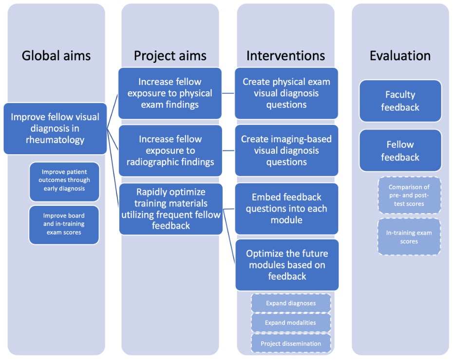 Figure 1: Project aims, interventions and evaluation. Future directions noted in lighter, dashed boxes.
.jpg) Figure 2: Process of module modification using a focus, feedback and action cycle.
Figure 2: Process of module modification using a focus, feedback and action cycle.
.jpg) Figure 3: Self-reported confidence levels before and after the module, measured through surveys using a 5-point Likert scale.
Figure 3: Self-reported confidence levels before and after the module, measured through surveys using a 5-point Likert scale.
To cite this abstract in AMA style:
Randhawa A, Nichols L, Kolfenbach J, Morcos M, Taylor S. I Know It When I See It! Improving Visual Diagnosis Education in Rheumatology Fellowship Training [abstract]. Arthritis Rheumatol. 2025; 77 (suppl 9). https://acrabstracts.org/abstract/i-know-it-when-i-see-it-improving-visual-diagnosis-education-in-rheumatology-fellowship-training/. Accessed .« Back to ACR Convergence 2025
ACR Meeting Abstracts - https://acrabstracts.org/abstract/i-know-it-when-i-see-it-improving-visual-diagnosis-education-in-rheumatology-fellowship-training/