
Session Information
Session Type: Poster Session (Monday)
Session Time: 9:00AM-11:00AM
Background/Purpose: One hypothesized link between cardiovascular disease and gout is the direct deposition of monosodium urate (MSU) crystals in atherosclerotic plaque. A 2018 ACR abstract used non-ECG-gated CT scans of the chest and neck in dual-energy mode to evaluate vascular MSU crystal deposition and reported that > 80% of patients with gout had vascular MSU deposition as well as some in healthy control patients, with no significant difference in the volume of MSU crystal deposition among gout and control patients. We reviewed 96 non-ECG gated DECT pulmonary angiograms (similar to the ACR abstract) to identify commonly encountered artifacts which could be misconstrued as true MSU deposition findings.
Methods: Consecutive patients with gout (ICD-10 code for gout and serum urate >6 mg/dL) who underwent non-ECG gated DECT pulmonary angiograms between 1/1/15 and 11/29/18 were identified using a clinical data repository. Age- and sex-matched controls were randomly chosen from a list of consecutive patients without gout who underwent the same imaging study. All images were acquired using dual-source, dual-energy CT scanners (Siemens Flash or Force DECT) and loaded into the postprocessing software (Syngo.via, Siemens, Germany) to apply an image-based two material decomposition algorithm to highlight MSU crystals in green. The images were reviewed by two radiologists experienced in interpreting DECT for MSU crystals. Presence of any green material in the vasculature was noted with its location for each patient, then further assessed with comparison to the grey-scale source images to determine if it was a true finding or artifact. Artifacts were classified into one of five categories: streak, misregistration due to motion, contrast mixing, foreign body, and noise. Three radiologists also reviewed two cases of ECG-gated coronary DECT scans.
Results: We identified 48 gout patients and 48 age- and sex-matched controls who had undergone 51 non ECG gated PE scans. The patients were mostly men (70.8%) with mean age 67 years. The majority of DECT scans, both of cases and controls, had green material in the vasculature (85% and 84%, respectively). The most common location of green material was in the superior vena cava or right atrium (81% in cases vs. 91% in controls), followed by the aorta (27% vs 47%, respectively) and coronary arteries (40% vs 21%, respectively). However, all green material in the vasculature were ultimately attributed to artifact with the most common types of artifacts being streak (Image 1) and misregistration due to motion (Image 2). However, images from the two gout patients using ECG-gated DECT scan showed coronary MSU deposits in both the grey-scale source images as well as dual energy impages, not meeting any of artifact criteria (Image 3).
Conclusion: Non-ECG gated Vascular DECT images are susceptible to various artifacts. Further investigation of this technology using ECG-gated CTs are warranted.

ACR 2019 DECT PE Image 1
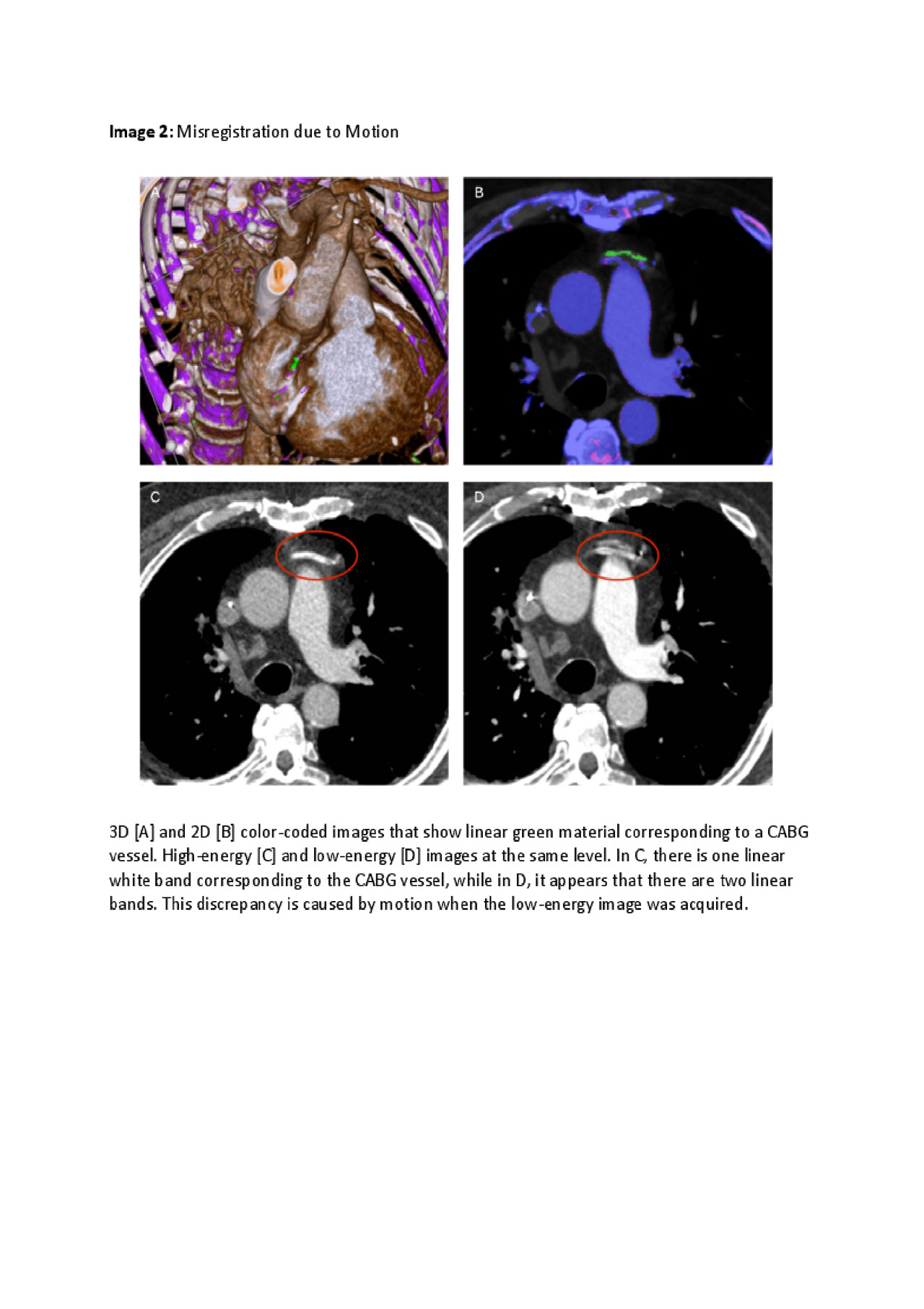
ACR 2019 DECT PE Image 2

ACR 2019 DECT PE Image 3
To cite this abstract in AMA style:
Yokose C, Eide S, Simeone F, Shojania K, Nicolaou S, Becce F, Choi H. Frequently Encountered Artifacts in Novel Application of Dual-Energy CT to Vascular Imaging: A Pilot Study [abstract]. Arthritis Rheumatol. 2019; 71 (suppl 10). https://acrabstracts.org/abstract/frequently-encountered-artifacts-in-novel-application-of-dual-energy-ct-to-vascular-imaging-a-pilot-study/. Accessed .« Back to 2019 ACR/ARP Annual Meeting
ACR Meeting Abstracts - https://acrabstracts.org/abstract/frequently-encountered-artifacts-in-novel-application-of-dual-energy-ct-to-vascular-imaging-a-pilot-study/