Session Information
Session Type: Poster Session (Sunday)
Session Time: 9:00AM-11:00AM
Background/Purpose: To evaluate the frequency and associations of osteoarthritis (KOA) features on knee ultrasound (KUS) in a community-based cohort study with radiographic and symptomatic data in the same knees.
Methods: A radiology technologist trained in standardized KUS imaging (SSG) scanned both knees in consecutive individuals enrolled in the Johnston County OA Project, using a written protocol. The KUS protocol included 7 views per knee: longitudinal and transverse suprapatellar in 30 degrees flexion (grading for effusion, gray scale synovitis and color power Doppler [CPD]), medial and lateral longitudinal (for osteophytes, meniscal damage, calcium deposition), maximally flexed suprapatellar transverse (for cartilage damage, calcium deposition) and posterior transverse (for popliteal cysts). Each set of images was scored using an atlas by 2 readers (previously shown to be reliable) whose scores were averaged. Radiographs (XR) were scored separately by an expert radiologist (JBR); all readers were blinded to other imaging and clinical data. Radiographic KOA (rKOA) was defined as a Kellgren-Lawrence grade (KLG) of 2 or more; osteophytes and joint space narrowing (JSN) were scored 0-3 using the OARSI atlas. Symptomatic KOA (sxKOA) was defined as rKOA with symptoms experienced in the same knee. Pain was assessed via the Knee Injury and OA Outcome Score (KOOS) pain subscale for each knee. We produced unadjusted Spearman correlations and additionally tested for nonzero correlation using the Cochran-Mantel-Haenszel statistic to describe associations with each KUS and XR feature and pain. All results shown are for right knees; left knees demonstrated similar patterns.
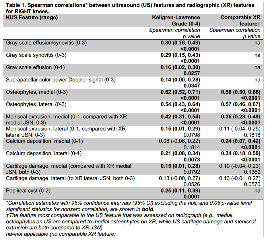
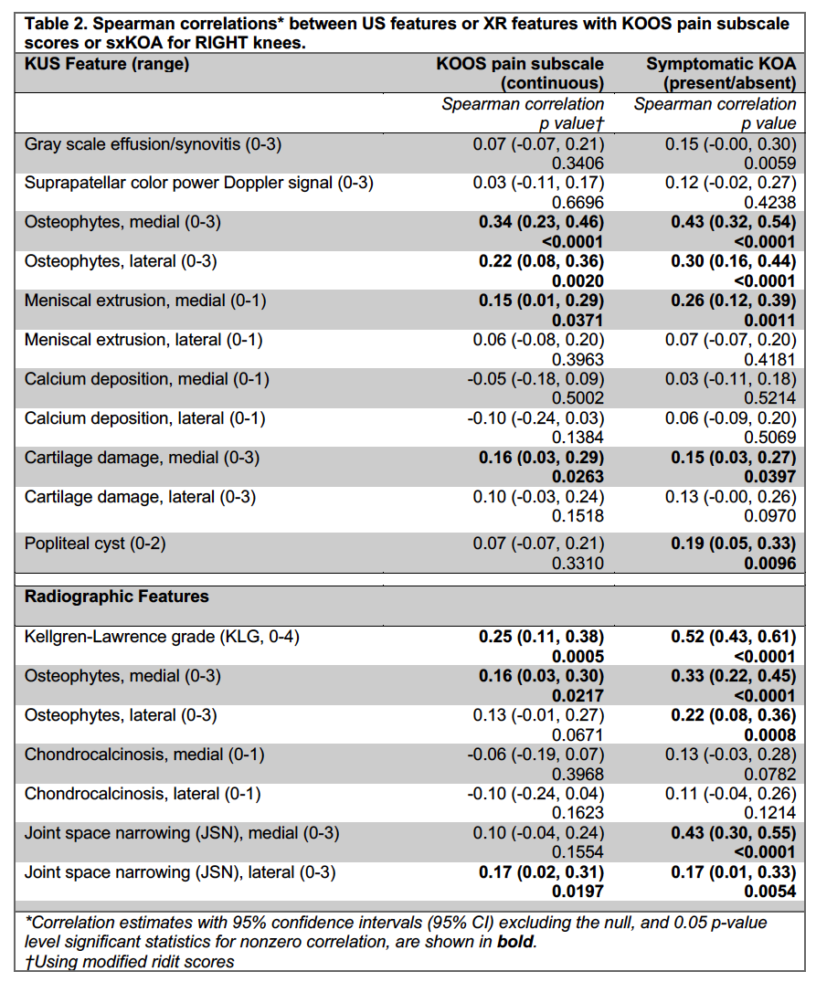
Results: Participants (n=203) had a mean (±SD) age of 73 ± 8 years and a mean BMI of 29.4 ± 7 kg/m2; about 1/3 were male and 1/3 were African-American. About a third of knees had symptoms and 5% had a history of knee injury. About half of knees met the above definition for rKOA, while almost a quarter met the sxKOA definition. The majority of knees had US evidence (score >0) of at least one of the following: effusion/synovitis, osteophytes, and/or cartilage damage (data not shown). Correlations between US and XR features are shown in Table 1. The strongest correlations were seen for osteophytes (r=0.6); similar correlations were seen between US osteophytes and XR KLG. Correlations for calcium deposition detected by each modality (r=0.2-0.3) were significant. Non-identical constructs such as medial US meniscal extrusion and XR JSN (r=0.4) were also significantly correlated. Medial XR JSN was more closely related to US meniscal extrusion (r=0.4) than to US cartilage damage (r=0.1). Osteophytes by US, compared with XR, had slightly stronger correlations with KOOS pain (Table 2). Medial meniscal extrusion and cartilage damage by US were significantly correlated with KOOS pain while medial JSN by XR was not; all three were correlated with presence of sxKOA.
Conclusion: US assessment of KOA is accessible and reliable and provides information complementary to XR; KUS may provide increased sensitivity for early KOA changes. Future work will further examine the associations between US and radiographic features, including effect modification by key covariates.
To cite this abstract in AMA style:
Yerich N, Alvarez C, Schwartz T, Savage-Guin S, Bakewell C, Kohler M, Lin J, Samuels J, Nelson A. Frequency of Ultrasound Features of Knee Osteoarthritis and Their Association with Radiographic Features and Symptoms in a Community-Based Cohort [abstract]. Arthritis Rheumatol. 2019; 71 (suppl 10). https://acrabstracts.org/abstract/frequency-of-ultrasound-features-of-knee-osteoarthritis-and-their-association-with-radiographic-features-and-symptoms-in-a-community-based-cohort/. Accessed .« Back to 2019 ACR/ARP Annual Meeting
ACR Meeting Abstracts - https://acrabstracts.org/abstract/frequency-of-ultrasound-features-of-knee-osteoarthritis-and-their-association-with-radiographic-features-and-symptoms-in-a-community-based-cohort/


