ACR Meeting Abstracts

ACR Meeting Abstracts

  • Meetings
    • ACR Convergence 2024
    • ACR Convergence 2023
    • 2023 ACR/ARP PRSYM
    • ACR Convergence 2022
    • ACR Convergence 2021
    • ACR Convergence 2020
    • 2020 ACR/ARP PRSYM
    • 2019 ACR/ARP Annual Meeting
    • 2018-2009 Meetings
    • Download Abstracts
  • Keyword Index
  • Advanced Search
  • Your Favorites
    • Favorites
    • Login
    • View and print all favorites
    • Clear all your favorites
  • ACR Meetings

Abstract Number: 1883

Elucidating Pain Mechanisms in Polyarticular Juvenile Idiopathic Arthritis Patients

Jaymin Upadhyay1, Diana Sibai 1, Robert Labadie 2, Jordan Lemme 3, Christine Sieberg 2, Kirsten Ecklund 4, David Borsook 4 and Robert Sundel 4, 1Boston Children's Hospital, Harvard Medical School, Boston, MA, 2Boston Children’s Hospital, Harvard Medical School, Boston, MA, 3Boston Children's Hospital, Harvard Medical School, Boston, 4Boston Children’s Hospital, Harvard Medical School, Boston

Meeting: 2019 ACR/ARP Annual Meeting

Keywords: inflammatory arthritis and magnetic resonance imaging (MRI), juvenile idiopathic arthritis (JIA), Neuroimaging, pain

  • Tweet
  • Click to email a link to a friend (Opens in new window) Email
  • Click to print (Opens in new window) Print
Session Information

Date: Monday, November 11, 2019

Title: 4M115: Pain Mechanisms – Basic & Clinical Science (1878–1883)

Session Type: ACR Abstract Session

Session Time: 4:30PM-6:00PM

Background/Purpose: Despite a broad range of available therapeutics, pain remains poorly treated in many juvenile idiopathic arthritis (JIA) patients1-2. For some patients, the level of pain is not commensurate with the magnitude of detectable joint pathology (i.e., synovitis). In order to elucidate pain mechanisms in JIA, we have investigated the functionality of CNS circuitry in polyarticular JIA (pJIA) individuals ( >5 affected joints) alongside peripheral joint pathology, markers of inflammation, subjective measures related to pain and clinical status. This strategy is projected to facilitate the identification of pain biomarker signatures and define mechanisms that underlie the observed pain-inflammation disconnect in JIA.

Methods: Consented, male and female pJIA patients (8-16 years) with and without pain were assessed using functional magnetic resonance imaging (fMRI), musculoskeletal MRI, Patient Reported Outcomes Measurement Information System (PROMIS) questionnaires, fluid markers informing on inflammation (i.e., CRP and ESR levels) and clinical status. All evaluated patients had either past or ongoing hand joint abnormalities. Patient enrollment is currently active.
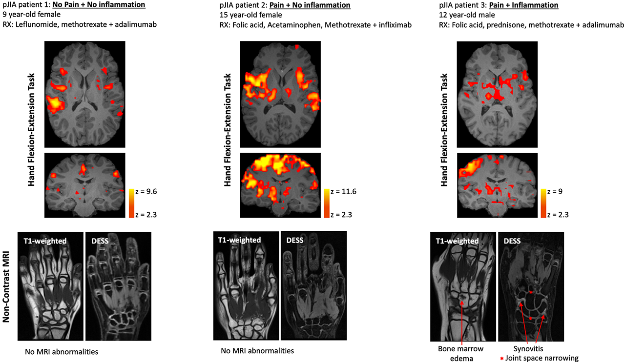
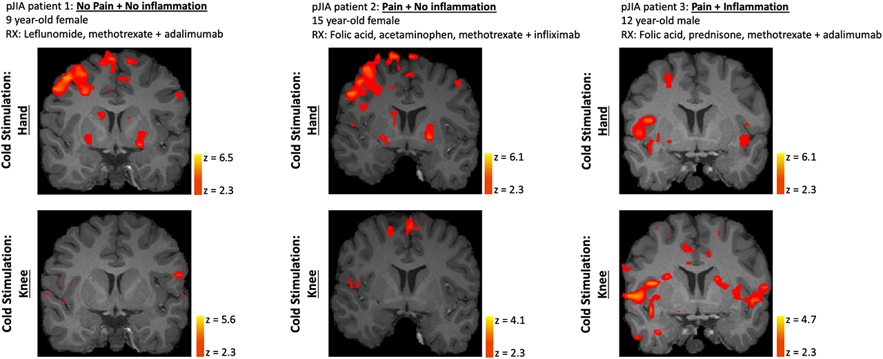
FMRI data collected during a hand motor task involved a visually cued, opening and closing of the right or left hand (depending on location of joint pathology or clinical pain) in a 30 second off/10 second on manner for 5 cycles. Evoked stimulation was subsequently performed by applying cold stimuli (10°C) to hand and knee joints also in a 30 second off/10 second on block design (Medoc LTD, Israel). Immediately following fMRI, patients underwent hand MRI (index hand only). All imaging was performed on a Siemens 3T scanner (Siemens, Germany). fMRI data was analyzed using FSL 6.0, while hand MRI was clinically evaluated by a musculoskeletal radiologist (KE). This study was approved by the Institutional Review Board at Boston Children’s Hospital.

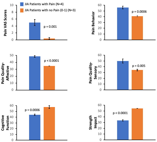
Results: Hand motor task fMRI showed differential CNS activation profiles amongst patients with No Pain + No inflammation, Pain + No Inflammation and Pain + Inflammation (Figure 1). Of these states, the pain without inflammation (or other pathology) sub-type yielded the most robust CNS activation. MRI abnormalities detected in the hand in Patient 3 consisted of (teno-)synovitis, bone marrow lesions and joint space narrowing. Cold stimuli yielded comparable responses across pain and inflammation conditions (Figure 2).  Interestingly, in the pJIA patient with current hand/wrist and past knee joint abnormalities, evoked CNS responses and pain scores to cold stimuli were similar at either site.  Finally, from PROMIS-based questionnaires (Figure 3), pJIA patients with pain reported a greater extent of loss of functions (cognition) and gain of functions (behaviors altered by pain).

Conclusion: For the first time, a neuroimaging-based assessment of pJIA patients was executed. This allowed for novel insight to be gained regarding potential neurobiological factors driving pain in pJIA. The current multidisciplinary datasets are suggestive of non-inflammatory (centralized or neuropathic) and inflammatory pain mechanisms at play in pJIA.

1. Giancane, Clin Exp Rheumatol. (2017)
2. Shiff, Pain. (2018) 

Figure 1: Evoked BOLD Response in pJIA Patients With and Without Pain During a Hand Motor Task. A. Single patient data showing CNS activity during a hand flexion and extension exercise. Statistical activation maps are depicted on axial -top row- and coronal -bottom row- planes on subject-specific, high-resolution anatomical MRI data. Patient 2 in particular reported pain and discomfort during the hand motor task. All pJIA patients had prior -Patient 1- or current -Patients 2 and 3- hand involvement in terms of clinical pain and/or inflammation. B. At the time neuroimaging evaluation, only one individual -Patient 3- had pathology detected in the hand as determined by non-contrast MRI. CRP and ESR levels were normal for Patients 1 and 2, but considered elevated in Patient 3 -CRP: 0.09 mg/dL and ESR: 24 mm/hr-.

Figure 2. Evoked CNS Response in pJIA Patients With and Without Pain During Unilateral Cold Stimulation of the Hand -Top Row- and Knee -Bottom Row-. Overall greater CNS activation was induced by cold stimulation of the dorsal side of the hand compared to the knee. Greater CNS activation for pJIA patient 3 for knee cold stimulation may have resulted from prior knee involvement. This patient reported pain during cold stimulation for both sites -hand and knee-. Statistical activation maps are depicted on subject-specific, high-resolution anatomical MRI data.

Figure 3. Group-Level Results Demonstrating the Impact of Pain in pJIA With and Without Pain. At the time of evaluation, pJIA patients reported clinical pain at a level of ~5 on a 0-10 scale. PROMIS-based questionnaires were used for all assessments, where higher T-score metrics represent more of the trait measured. The presence of pain in pJIA negatively impacted -loss of function- domains such as cognitive ability and strength. Two-tailed t-tests were performed for all comparisons.


Disclosure: J. Upadhyay, None; D. Sibai, None; R. Labadie, None; J. Lemme, None; C. Sieberg, None; K. Ecklund, None; D. Borsook, Biogen, 5; R. Sundel, None.

To cite this abstract in AMA style:

Upadhyay J, Sibai D, Labadie R, Lemme J, Sieberg C, Ecklund K, Borsook D, Sundel R. Elucidating Pain Mechanisms in Polyarticular Juvenile Idiopathic Arthritis Patients [abstract]. Arthritis Rheumatol. 2019; 71 (suppl 10). https://acrabstracts.org/abstract/elucidating-pain-mechanisms-in-polyarticular-juvenile-idiopathic-arthritis-patients/. Accessed .
  • Tweet
  • Click to email a link to a friend (Opens in new window) Email
  • Click to print (Opens in new window) Print

« Back to 2019 ACR/ARP Annual Meeting

ACR Meeting Abstracts - https://acrabstracts.org/abstract/elucidating-pain-mechanisms-in-polyarticular-juvenile-idiopathic-arthritis-patients/

Advanced Search

Your Favorites

You can save and print a list of your favorite abstracts during your browser session by clicking the “Favorite” button at the bottom of any abstract. View your favorites »

All abstracts accepted to ACR Convergence are under media embargo once the ACR has notified presenters of their abstract’s acceptance. They may be presented at other meetings or published as manuscripts after this time but should not be discussed in non-scholarly venues or outlets. The following embargo policies are strictly enforced by the ACR.

Accepted abstracts are made available to the public online in advance of the meeting and are published in a special online supplement of our scientific journal, Arthritis & Rheumatology. Information contained in those abstracts may not be released until the abstracts appear online. In an exception to the media embargo, academic institutions, private organizations, and companies with products whose value may be influenced by information contained in an abstract may issue a press release to coincide with the availability of an ACR abstract on the ACR website. However, the ACR continues to require that information that goes beyond that contained in the abstract (e.g., discussion of the abstract done as part of editorial news coverage) is under media embargo until 10:00 AM ET on November 14, 2024. Journalists with access to embargoed information cannot release articles or editorial news coverage before this time. Editorial news coverage is considered original articles/videos developed by employed journalists to report facts, commentary, and subject matter expert quotes in a narrative form using a variety of sources (e.g., research, announcements, press releases, events, etc.).

Violation of this policy may result in the abstract being withdrawn from the meeting and other measures deemed appropriate. Authors are responsible for notifying colleagues, institutions, communications firms, and all other stakeholders related to the development or promotion of the abstract about this policy. If you have questions about the ACR abstract embargo policy, please contact ACR abstracts staff at [email protected].

Wiley

  • Online Journal
  • Privacy Policy
  • Permissions Policies
  • Cookie Preferences

© Copyright 2025 American College of Rheumatology